. 2021 Sep 17;13(4):10.1088/1758-5090/ac23ae.
doi: 10.1088/1758-5090/ac23ae.

miRNA induced co-differentiation and cross-talk of adipose tissue-derived progenitor cells for 3D heterotypic pre-vascularized bone formation

Affiliations

miRNA induced co-differentiation and cross-talk of adipose tissue-derived progenitor cells for 3D heterotypic pre-vascularized bone formation

Nazmiye Celik et al. Biofabrication. .

Abstract

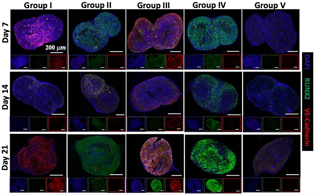
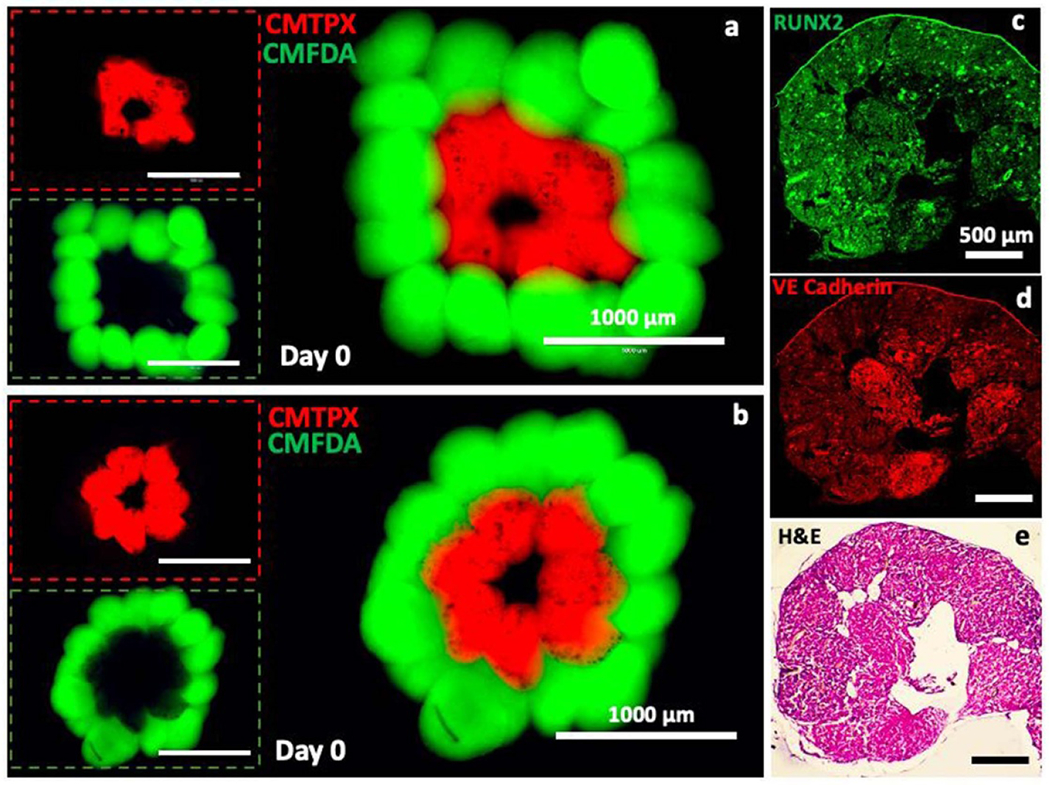
Engineered bone grafts require a vascular network to supply cells with oxygen, nutrients and remove waste. Using heterotypic mature cells to create these graftsin vivohas resulted in limited cell density, ectopic tissue formation and disorganized tissue. Despite evidence that progenitor cell aggregates, such as progenitor spheroids, are a potential candidate for fabrication of native-like pre-vascularized bone tissue, the factors dictating progenitor co-differentiation to create heterotypic pre-vascularized bone tissue remains poorly understood. In this study, we examined a three-dimensional heterotypic pre-vascularized bone tissue model, using osteogenic and endotheliogenic progenitor spheroids induced by miR-148b and miR-210 mimic transfection, respectively. Spheroids made of transfected cells were assembled into heterotypic structures to determine the impact on co-differentiation as a function of micro-RNA (miRNA) mimic treatment group and induction time. Our results demonstrated that miRNAs supported the differentiation in heterotypic structures, and that developing heterotypic structures is determined in part by progenitor maturity, as confirmed by gene and protein markers of osteogenic and endotheliogenic differentiation and the mineralization assay. As a proof of concept, miRNA-transfected spheroids were also bioprinted using aspiration-assisted bioprinting and organized into hollow structures to mimic the Haversian canal. Overall, the presented approach could be useful in fabrication of vascularized bone tissue using spheroids as building blocks.

Keywords: bioprinting; bone; miRNA; pre-vascularization; spheroid assembly; stem cells.


Conflict of interest statement

Conflict of interest

The authors declare no competing interests.

Figures

Figure 1.
Figure 1.
A schematic diagram describing biofabrication of doublet structures using miR-210 and miR-148b transfected spheroids. (Note: BM stands for basal medium and OM stands for osteogenic medium.)
Figure 2.
Figure 2.
Gene expression levels for Group I (transfected with miR-210), Group II (transfected with miR-148b), Group III (transfected with miR-148b and miR-210), and Group IV (positive control in OM medium) doublets normalized to Group V (negative control in basal medium) doublets for osteogenic markers: (a) RUNX2, (b) Col-1, (c) OSTERIX, and (d) BSP (n = 3; p* < 0.05; p** < 0.01; p*** < 0.001; p**** < 0.0001).
Figure 3.
Figure 3.
Gene expression levels for Group I (transfected with miR-210), Group II (transfected with miR-148b), Group III (transfected with miR-148b and miR-210), and Group IV (positive control in OM medium) doublets normalized to Group V (negative control in basal medium) doublets for endotheliogenic markers: (a) PECAM-1, and (b) ANGPT-1 (n = 3; p* < 0.05; p** < 0.01).
Figure 4.
Figure 4.
Immunostaining (DAPI in blue, RUNX2 in green, and VE-cadherin in red) images of Strategy-1 (2 d transfection period) doublets of Group I (transfected with miR-210), Group II (transfected with miR-148b), Group III (transfected with miR-148b and miR-210), Group IV (positive control in OM medium), and Group V (negative control in basal medium) at Days 7, 14, and 21. Scale bars in insets correspond to 200 μm.
Figure 5.
Figure 5.
Immunostaining (DAPI in blue, RUNX2 in green, and VE-cadherin in red) images of Strategy-2 (14 d transfection period) spheroids of Group I (transfected with miR-210), Group II (transfected with miR-148b), Group III (transfected with miR-148b and miR-210), Group IV (positive control in OM medium), and Group V (negative control in basal medium) at Days 7, 14, and 21. Scale bars in insets correspond to 200 μm.
Figure 6.
Figure 6.
Osteoimage staining of assembled doublet structures for (a) Strategy-1 (2 d transfection period) and (b) Strategy-2 (14 d transfection period). Quantitative intensity analysis of osteoimages for (c) Strategy-1 and (d) Strategy-2 (n = 3; p* < 0.05; p** < 0.01; p*** < 0.001). Group I (transfected with miR-210), Group II (transfected with miR-148b), Group III (transfected with miR-148b and miR-210), Group IV (positive control in OM medium), and Group V (negative control in basal medium) at Days 7, 14, and 21.
Figure 7.
Figure 7.
H&E images of doublet structures of (a) Strategy-1 (2 d transfection period) and (b) Strategy-2 (14 d transfection period) for Group I (transfected with miR-210), Group II (transfected with miR-148b), Group III (transfected with miR-148b and miR-210), Group IV (positive control in OM medium), and Group V (negative control in basal medium) at Days 7, 14, and 21.
Figure 8.
Figure 8.
Characterization of spheroid fusion in doublets. For Strategy-1 (2 d transfection period), (a) representative light microscopy images of fusing spheroids in assembled doublet structures over the 21 d timeframe and corresponding parameters including (b) doublet length (μm), (c) contact length (μm), (d) intersphere angle (°), and (e) doublet width (μm). For Strategy-2 (14 d transfection period), (f) representative light microscopy images of fusing spheroids in assembled doublet structures over the 21 d timeframe and corresponding parameters including (g) doublet length (μm), (h) contact length (μm), (i) intersphere angle (°), and (j) doublet width (μm). (k) A schematic showing morphological parameters measured during fusion.
Figure 9.
Figure 9.
Representation and characterization of the Haversian canal model fabricated using the aspiration-assisted bioprinting technique. (a), (b) Bioprinted structures consisted of ADSCs spheroids labeled with CellTracker™ green CMFDA dye and CellTracker™ Red CMTPX dye post bioprinting. Immunoimages, showing (c) RUNX2 and (d) VE-cadherin, and (e) a H&E image of a bioprinted structure at Day 14 post bioprinting.

References

    1. Soucacos PN, Kokkalis ZT, Piagkou M and Johnson EO 2013. Vascularized bone grafts for the management of skeletal defects in orthopaedic trauma and reconstructive surgery Injury 44 S70–5 - PubMed
    1. Amini AR, Laurencin CT and Nukavarapu SP 2012. Bone tissue engineering: recent advances and challenges Crit. Rev. Biomed. Eng 40 363–408 - PMC - PubMed
    1. Cai X, Yang F, Yan X, Yang W, Yu N, Oortgiesen DAW, Wang Y, Jansen JA and Walboomers XF 2015. Influence of bone marrow-derived mesenchymal stem cells pre-implantation differentiation approach on periodontal regeneration in vivo J. Clin. Periodontol 42 380–9 - PubMed
    1. Kuhn LT et al. 2014. Developmental-like bone regeneration by human embryonic stem cell-derived mesenchymal cells Tissue Eng. A 20 365–77 - PMC - PubMed
    1. Eppler SM, Combs DL, Henry TD, Lopez JJ, Ellis SG, Yi J-H, Annex BH, McCluskey ER and Zioncheck TF 2002. A target-mediated model to describe the pharmacokinetics and hemodynamic effects of recombinant human vascular endothelial growth factor in humans Clin. Pharmacol. Ther 72 20–32 - PubMed

Publication types