. 2021 Sep 3;21(1):990.
doi: 10.1186/s12885-021-08357-8.

Head and neck squamous cell carcinoma cell lines have an immunomodulatory effect on macrophages independent of hypoxia and toll-like receptor 9

Affiliations

Head and neck squamous cell carcinoma cell lines have an immunomodulatory effect on macrophages independent of hypoxia and toll-like receptor 9

Tamiko Ishizu et al. BMC Cancer. .

Abstract

Background: A low tissue oxygen level, < 1% O2, is a typical characteristic inside of solid tumors in head and neck cancer (HNSCC) affecting a wide array of cell populations, such as macrophages. However, the mechanisms of how hypoxia influences macrophages are not yet fully elucidated. Our research aimed to study the effect of soluble mediators produced by hypoxic cancer cells on macrophage polarization. Furthermore, we studied the effect of a hypoxic microenvironment on the expression of tumorigenic toll-like receptor 9 (TLR9) and the consecutive macrophage polarization.

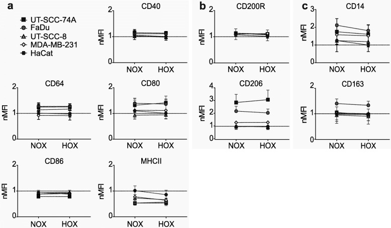
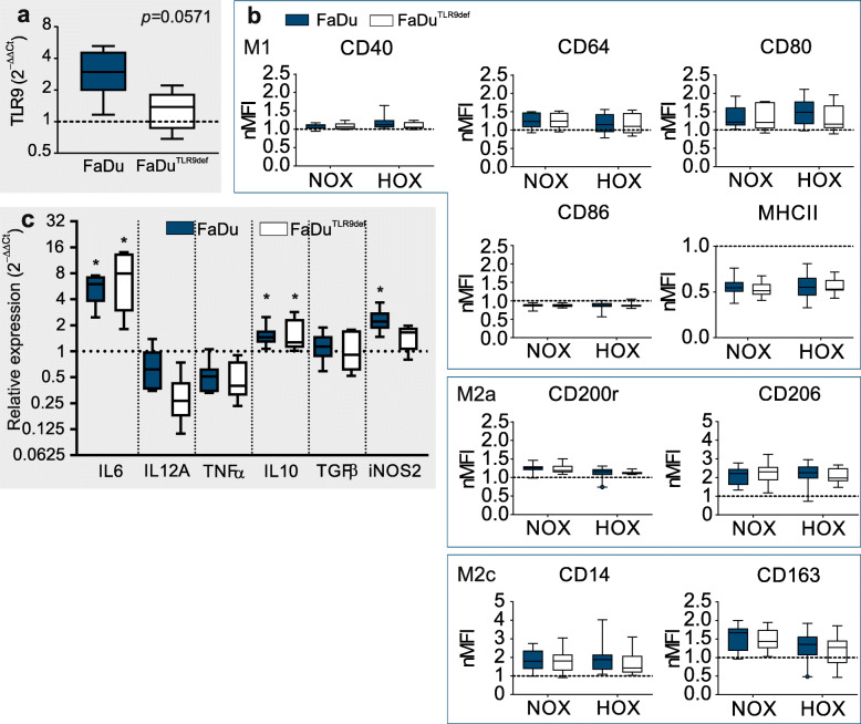
Methods: Conditioned media (CMNOX or CMHOX) from cell lines UT-SCC-8, UT-SCC-74A, FaDu, MDA-MB-231 and HaCat cultured under normoxic (21% O2) and hypoxic (1% O2) conditions were used to polarize human monocyte-derived macrophages. Macrophage polarization was measured by flow cytometry and the production of cytokine mRNA using Taqman qPCR. To study the role of TLR9 in macrophage polarization, the lentiviral CRISPR/Cas9 method was used to establish a stable FaDuTLR9def clone.

Results: Our results demonstrate that the soluble mediators produced by the cancer cells under normoxia polarize macrophages towards a hybridized M1/M2a/M2c phenotype. Furthermore, the results suggest that hypoxia has a limited role in altering the array of cancer-produced soluble factors affecting macrophage polarization and cytokine production. Our data also indicates that increased expression of TLR9 due to hypoxia in malignant cells does not markedly influence the polarization of macrophages. TLR9 transcriptional response to hypoxia is dissimilar to a HIF1-α-regulated LDH-A. This may indicate a context-dependent expression of TLR9 under hypoxia.

Conclusions: HNSCC cell lines affect both macrophage activity (polarization) and functionality (cytokines), but with exception to iNOS expression, the effects appear independent of hypoxia and TLR9.

Keywords: Anti-cancer immunomodulation; Head and neck squamous cell carcinoma; Hypoxia; Immune evasion; Immunoediting; Innate immune response; Macrophage polarization; TLR9.


Conflict of interest statement

The authors declare that they have no competing interests.

Figures

Fig. 1
Fig. 1
Changes in the relative phenotypical features of non-activated macrophages post conditioned media treatment (a) Outline of the conditioned media (CM) -experiment. (b, c, d) Following incubation with CMNOX for two days, macrophage (MΦ) polarization was assessed with flow cytometry. Panels b, c, and d illustrate M1, M2a and M2c polarization markers, respectively. UT-SCC-8 and UT-SCC-74A are abbreviated here as SCC-8 and SCC-74A. Box plots represent a comparison of normalized MFI (nMFI) values against NA MΦ (dotted line), error bars 95% CI. Experiments were repeated a minimum of 5 times. *p ≤ 0.05 vs. NA MΦ was considered statistically significant (the exact p-values can be found in Additional file F7b)
Fig. 2
Fig. 2
Expression of macrophage surface markers post-incubation with hypoxia-conditioned media (CMHOX). Conditioned media collected after 24 h exposure was used to study macrophage polarization. Panel (a) shows the resulting change in M1, (b) in M2a and (c) in M2c marker expression. The dotted line represents non-activated macrophages. Results from a minimum of 5 independent experiments are shown as geometric mean of normalized MFI (nMFI) with 95% CI
Fig. 3
Fig. 3
Lactate dehydrogenase A expression under hypoxia compared with normoxia. Succesful hypoxia in cancer cells was verified by measuring hypoxia-inducible factor 1α- regulated lactate dehydrogenase A (LDH-A) expression after 24 h exposure to hypoxia (normoxia, dotted line). Data from a minimum of 4 independent experiments, except HaCat (n = 3), is shown as box plots with 95% CI
Fig. 4
Fig. 4
Cytokine mRNA expression in macrophages (MΦ) exposed to conditioned media (CM) Following 2-day exposure to either polarizing cytokines or conditioned media from FaDu cells collected under normoxia (CMNOX) or hypoxia (CMHOX), MΦ mRNA was extracted and the cytokine expression profile was assessed. Panel (a) shows cytokines produced by M1, M2a, and M2c polarization controls. Panel (b) shows the cytokine mRNA expression after treatment with CMNOX or CMHOX from parental FaDu cells. Experiments were repeated a minimum of 5 times, except in the control groups (n ≥ 2). *p ≤ 0.05 vs. NA MΦ (dotted line), the exact p-values can be found in Additional file 5
Fig. 5
Fig. 5
The influence of FaDu and FaDuTLR9def conditioned media on macrophage polarization and mRNA cytokine expression. (a) After 48 h of exposure to hypoxia, mRNA from parental FaDu and FaDuTLR9def cells was extracted to assess their toll-like receptor 9 (TLR9) expression (normoxia, dotted line). p = 0.0571, n = 4. (b) Non-activated macrophages (NA MΦ) (dotted line) were treated with conditioned media (CM) for 2 days prior assessing their polarization phenotype with flow cytometry. Panel (c) illustrates cytokine mRNA expression from MΦ after treatment with CM from hypoxic parental FaDu or FaDuTLR9def cells. Results are shown as box plots with 95% CI. The dotted line represents NA MΦ. Experiments were repeated a minimum of 5 times. NA vs. CMHOX-treated MΦ was considered statistically significant when *p ≤ 0.05. The exact p-values can be found in Additional File 5b

References

    1. Wynn TA, Chawla A, Pollard JW. Macrophage biology in development, homeostasis and disease. Nature. 2013;496(7446):445–455. doi: 10.1038/nature12034. - DOI - PMC - PubMed
    1. Tarique AA, Logan J, Thomas E, Holt PG, Sly PD, Fantino E. Phenotypic, functional, and plasticity features of classical and alternatively activated human macrophages. Am J Respir Cell Mol Biol. 2015;53(5):676–688. doi: 10.1165/rcmb.2015-0012OC. - DOI - PubMed
    1. Martinez FO, Gordon S. The M1 and M2 paradigm of macrophage activation: time for reassessment. F1000Prime Rep. 2014;6:1–13. doi: 10.12703/P6-13. - DOI - PMC - PubMed
    1. Quatromoni JG, Eruslanov E. Tumor-associated macrophages: function, phenotype, and link to prognosis in human lung cancer. Am J Transl Res. 2012;4(4):376–389. - PMC - PubMed
    1. Lurier EB, Dalton D, Dampier W, Raman P, Nassiri S, Ferraro NM, Rajagopalan R, Sarmady M, Spiller KL. Transcriptome analysis of IL-10-stimulated (M2c) macrophages by next-generation sequencing. Immunobiology. 2017;222(7):847–856. doi: 10.1016/j.imbio.2017.02.006. - DOI - PMC - PubMed

MeSH terms