. 2021 Sep 3;21(1):335.
doi: 10.1186/s12883-021-02366-7.

Impairment of the visuospatial working memory in the patients with Parkinson's Disease: an fMRI study

Affiliations

Impairment of the visuospatial working memory in the patients with Parkinson's Disease: an fMRI study

Shoji Kawashima et al. BMC Neurol. .

Abstract

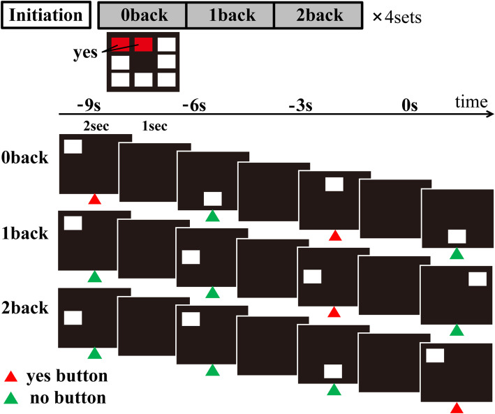
Background: Mild cognitive impairment (MCI) is a common symptom in the patients with Parkinson's disease (PD). The characteristics of cognitive impairment in PD are executive function (including working memory) and visuo-perceptual processing. The visuospatial n-back test has the merit of minimizing the influence of educational biases involved in the verbal n-back test. Furthermore, it can assess both visuospatial recognition and working memory in a single test.

Methods: We aimed to clarify the advantage of the visuospatial n-back test as a tool for detecting impairments of working memory in PD. We enrolled 28 right-handed patients with PD (18 males, 10 females) and 12 age-matched healthy controls (HC; 7 males, 5 females). Thirteen patients were classified as MCI (PD-MCI), and 15 as cognitively normal PD (PD-CN). Using functional MRI (fMRI), we explored the specific brain regions associated with the performance of the n-back test in the PD-MCI, PD-CN, and HC groups. The 0-back test assesses visuospatial recognition, while the 1-back and 2-back tests assess visuospatial working memory. Group comparisons were performed for three loads of this test.

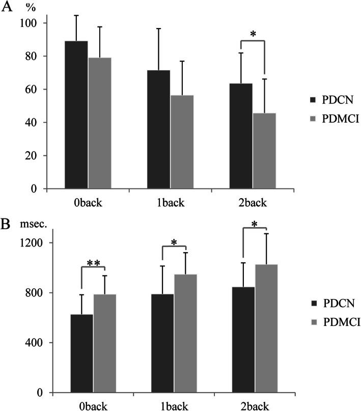
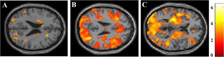
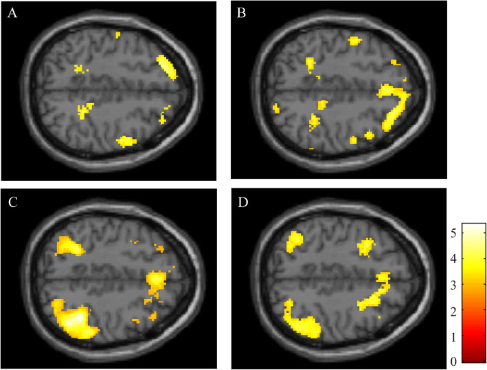
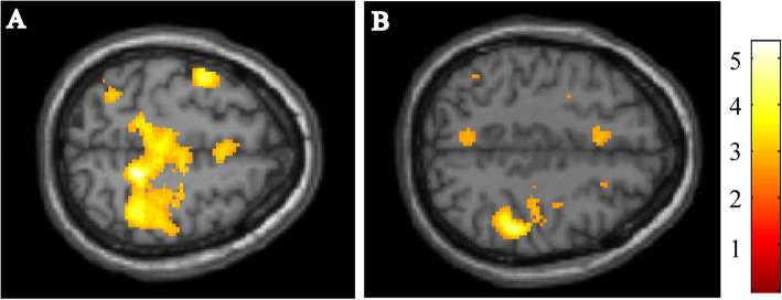
Results: Patients with PD performed significantly worse in terms of the correct answer rates of all n-back tests compared with HC. fMRI analyses performed during the 2-back test revealed reduced activation in the bilateral dorsolateral prefrontal cortex, middle frontal gyrus (MFG), and parietal lobule in the PD group compared with the HC group. In contrast, the fMRI result during the 0-back test showed only a marginal difference in the frontal lobe. On comparisons of task performance between the PD-MCI and PD-CN groups, we found that the correct answer rate in the 2-back test was lower in the PD-MCI group than in the PD-CN group. However, scores of the 0-back and 1-back tests were not significantly different between the two groups. The fMRI findings revealed that activations within the middle frontal gyrus (MFG) and inferior parietal lobule (IPL) during the 2-back test were reduced in the patients with PD-MCI when compared to those with PD-CN.

Conclusions: This study reports reduced activation of the MFG and IPL in patients with PD-MCI. These regions may be associated with the pathophysiology of working memory impairment in patients with PD, which involves fronto-striatal network dysfunction.

Keywords: Mild cognitive impairment; Parkinson’s disease; functional MRI; n-back test; working memory.


Conflict of interest statement

The authors declare that they have no competing interests.

Figures

Fig. 1
Fig. 1
Experimental protocol. The figure shows the protocol of n-back task used in this study. To detect the activated brain regions associated with the visuospatial n-back task, we used a blocked design fMRI, alternating n-back task conditions and rest conditions. In n-back task conditions, the white square was presented for 2 s at random in 1 of 8 possible locations on screen (exposure time), and black screen was presented for 1 s after the presentation of stimuli (interstimuli interval). Therefore, the consecutive stimuli were presented every 3 s. In the 0-back test, the subjects were instructed to press the left button with their index finger when the white square was presented in predetermined locations. They were instructed to press the right button when the stimulus appeared in any other location. In the 1-back test, the subjects were instructed to press the left button when the stimulus presented in the same location as the previous one. In the 2-back test, they were instructed to press the left button whenever a stimulus was presented in the same location as the two trials previous. When the stimulus was presented in any other location during the 1-back and 2-back test, the patients were instructed to press the right button with the middle finger
Fig. 2
Fig. 2
Performance of the visuospatial n-back test in the patients with PD. A. The figure shows rates of correct answers for the patients with PD-CN and PD-MCI. The scores on the 0-back test did not differ significantly between groups. The scores on the 2-back test of PD-MCI were significantly lower than that of PD-CN. B. The figure shows the response time to press correct button. The patients with PD-MCI took significantly longer time to respond in all task conditions. * P < 0.05, ** P < 0.01
Fig. 3
Fig. 3
fMRI analyses comparing the patients with PD and HC. The figure shows the results for the 0-back test (A), 2-back test (B) and subtracting the activated regions in the 0-back test from those in the 2-back test (C). The coloured regions indicate significantly lower brain activation in patients with PD, as compared with HC. The colour-bar represents t values as reference. All the images presented at P < 0.01 (uncorrected) with cluster size > 50 voxels in analysis. (A) The fMRI result during the 0-back test showed only a marginal difference in frontal lobe. (B) The result of the 2-back test showed the reduced activation in the bilateral dorsolateral prefrontal cortex, middle frontal gyrus, inferior parietal lobule, and superior parietal lobule. (C) The result of 2-back versus 0-back condition presented reduction of the activation in the bilateral prefrontal cortex, occipital cortex, and bilateral putamen
Fig. 4
Fig. 4
fMRI analyses comparing PD-MCI and PD-CN. These images show the results for the 0-back test (A), 1-back test (B), 2-back test (C), and subtracting the activated regions in the 0-back test from those in the 2-back test (2-back versus 0-back condition) (D). The coloured regions indicate significantly lower brain activation in PD-MCI, as compared with PD-CN. The colour-bar represents t values as reference. All the images presented at P < 0.01 (uncorrected) with cluster size > 50 voxels in analysis
Fig. 5
Fig. 5
fMRI correlation analyses for the patients with PD-MCI. The figure shows the results of correlations between the scores on the n-back test and task-related activation in the PD-MCI group. The colour-bar represents t values as reference. All the images presented at P < 0.001 (uncorrected) with cluster size > 50. A. The image shows the correlation between the scores of the 1-back test and activation of the 1-back test. B. The image shows the correlation between the scores of the 2-back test and activation of the 2-back test

References

    1. Emre M, Aarsland D, Brown R, Burn D, Duyckaerts C, Mizuno Y, et al. Clinical diagnostic criteria for dementia associated with Parkinson’s disease. Mov Disord. 2007;22(12):1689–707. doi: 10.1002/mds.21507. - DOI - PubMed
    1. Litvan I, Goldman JG, Troster AI, Schmand BA, Weintraub D, Petersen RC, et al. Diagnostic criteria for mild cognitive impairment in Parkinson’s disease. Movement Disorder Society Task Force guidelines. Mov Disord. 2012;27(3):349–56. doi: 10.1002/mds.24893. - DOI - PMC - PubMed
    1. Petersen RC, Roberts RO, Knopman DS, Boeve BF, Geda YE, Ivnik RJ, et al. Mild cognitive impairment: ten years later. Arch Neurol. 2009;66(12):1447–55. doi: 10.1001/archneurol.2009.266. - DOI - PMC - PubMed
    1. Pedersen KF, Larsen JP, Tysnes OB, Alves G. Natural course of mild cognitive impairment in Parkinson disease: A 5-year population-based study. Neurology. 2017;88(8):767–774. doi: 10.1212/WNL.0000000000003634. - DOI - PubMed
    1. Lee JE, Cho KH, Song SK, Kim HJ, Lee HS, Sohn YH, et al. Exploratory analysis of neuropsychological and neuroanatomical correlates of progressive mild cognitive impairment in Parkinson’s disease. JNNP. 2013;85(1):7–16. - PubMed