Case Reports
. 2021 Sep 3;14(9):e244023.
doi: 10.1136/bcr-2021-244023.

Saline contrast echocardiography complements cardiac interventions in neonates with transposition of great arteries and abnormal ductus venosus anatomy

Affiliations
Case Reports

Saline contrast echocardiography complements cardiac interventions in neonates with transposition of great arteries and abnormal ductus venosus anatomy

Monika Kantilal Kotecha et al. BMJ Case Rep. .

Abstract

We present a rare case of premature low birthweight neonate with right diaphragmatic hernia and transposition of great vessels requiring balloon atrial septostomy. Congenital diaphragmatic hernia poses a unique challenge to umbilical venous catheterisation. Based on the radiographic position of umbilical vein catheter, umbilical venous cannulation was attempted; however, the catheter could not be navigated to the right atrium. Saline contrast echocardiography was used to delineate the abnormal umbilical and ductus venosus drainage. Eventually, the procedure was successfully completed via the femoral venous approach. We emphasise the importance of defining ductus venosus anatomy and umbilical venous drainage using a simple tool like saline contrast echocardiography before performing catheterisation using the umbilical venous access in such cases.

Keywords: clinical diagnostic tests; congenital disorders; interventional cardiology; neonatal intensive care; ultrasonography.


Conflict of interest statement

Competing interests: None declared.

Figures
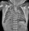
Figure 1
Figure 1
Anteroposterior chest radiograph showing the tip of umbilical venous catheter (black arrow) to the right of T9–T10 vertebral bodies.
Figure 2
Figure 2
Echocardiographic images of umbilical venous catheter. (A) Subcostal echocardiographic image showing umbilical venous catheter and the RA. (B) Subcostal echocardiographic image showing RA, LA, guide wire in RA (white arrow) and umbilical venous catheter (black arrow) posterior to the LA. LA, left atrium; RA, right atrium.
Figure 3
Figure 3
Saline contrast study. (A) Subcostal echocardiographic image showing hepatic vein (arrow) joining the IVC, which then enters the RA. (B) Subcostal echocardiographic image after saline contrast injection showing microbubbles in hepatic vein (arrow) and not in the IVC. IVC, inferior vena cava; RA, right atrium.
Figure 4
Figure 4
Illustration of umbilical venous drainage in normal neonate and altered anatomy of ductus venosus in right congenital diaphragmatic hernia. (A) Schematic illustration for normal drainage of umbilical vein. Umbilical venous catheter passes from the umbilical vein to the ductus venosus to enter the right atrium. (B) Schematic illustration showing the course of umbilical venous catheter into the right portal vein in case of right diaphragmatic hernia with liver herniation. Note the horizontal course of ductus venosus into the inferior vena cava.

References

    1. Sakurai M, Donnelly LF, Klosterman LA, et al. . Congenital diaphragmatic hernia in neonates: variations in umbilical catheter and enteric tube position. Radiology 2000;216:112–6. 10.1148/radiology.216.1.r00jl13112 - DOI - PubMed
    1. Richards DS, Kays DM. Fetal umbilical vein deviation in congenital diaphragmatic hernia. J Ultrasound Med 2013;32:263–8. 10.7863/jum.2013.32.2.263 - DOI - PubMed
    1. Chang PT, Taylor GA. Umbilical venous catheter malposition and errors in interpretation in newborns with Bochdalek hernia. Pediatr Radiol 2015;45:982–8. 10.1007/s00247-014-3275-7 - DOI - PubMed
    1. Montalva L, Lauriti G, Zani A. Congenital heart disease associated with congenital diaphragmatic hernia: a systematic review on incidence, prenatal diagnosis, management, and outcome. J Pediatr Surg 2019;54:909–19. 10.1016/j.jpedsurg.2019.01.018 - DOI - PubMed
    1. Simpson JM, Moore P, Teitel DF. Cardiac catheterization of low birth weight infants. Am J Cardiol 2001;87:1372–7. 10.1016/S0002-9149(01)01555-7 - DOI - PubMed

Publication types

MeSH terms