. 2022 Jun;43(6):1360-1371.
doi: 10.1038/s41401-021-00759-5. Epub 2021 Sep 3.

Metformin promotes microglial cells to facilitate myelin debris clearance and accelerate nerve repairment after spinal cord injury

Affiliations

Metformin promotes microglial cells to facilitate myelin debris clearance and accelerate nerve repairment after spinal cord injury

Yan-Qing Wu et al. Acta Pharmacol Sin. 2022 Jun.

Abstract

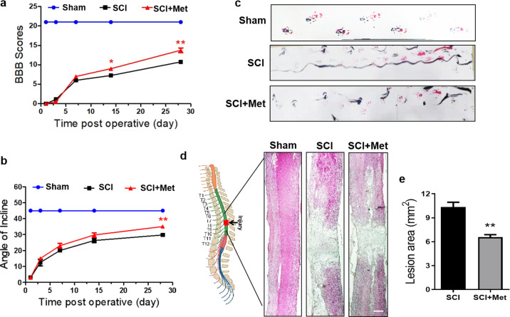
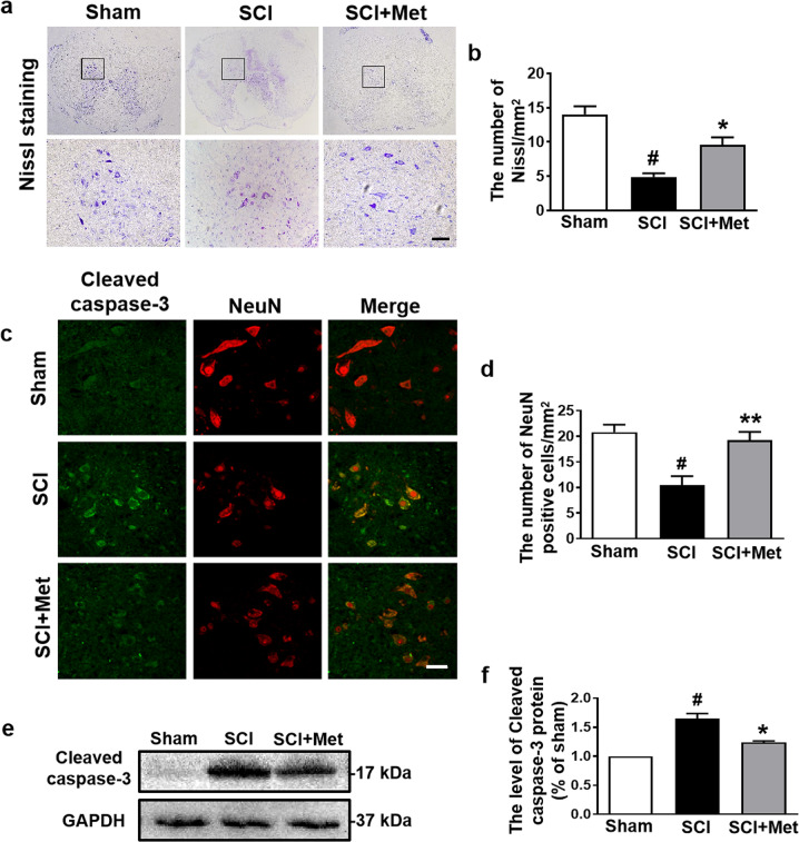
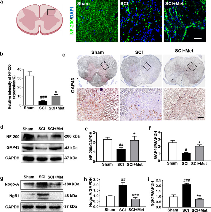
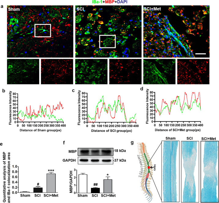
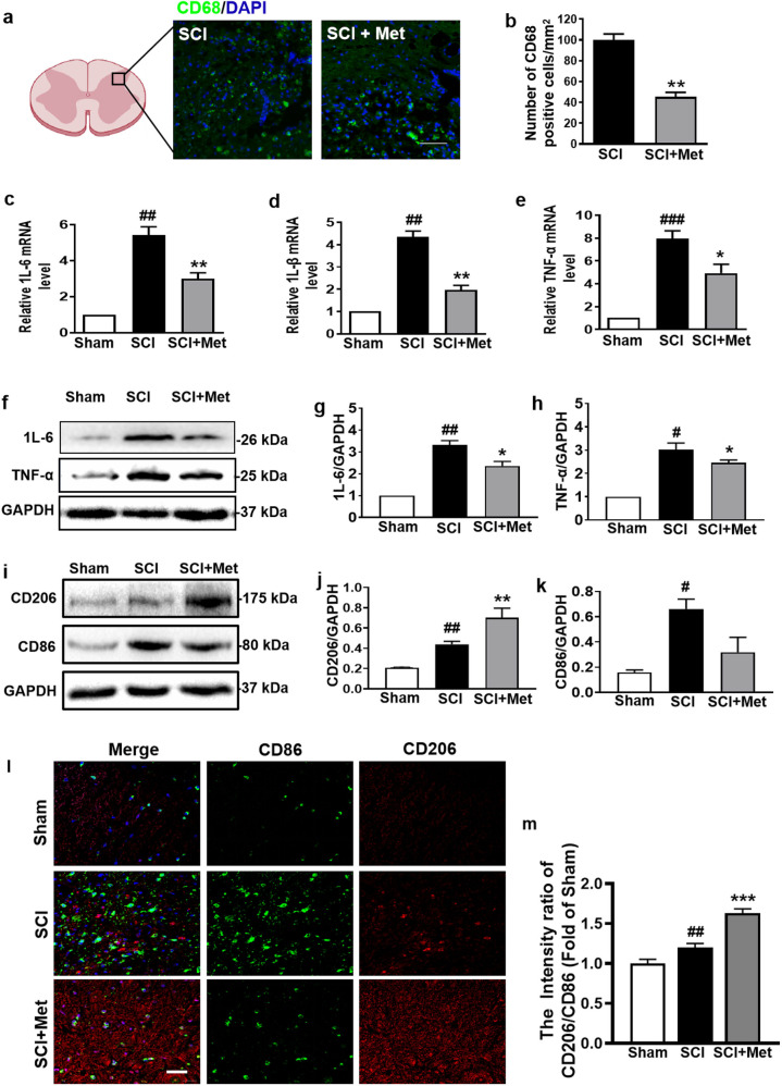
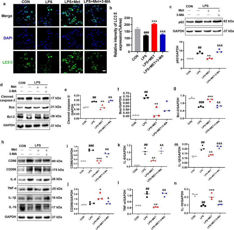
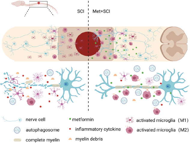
Spinal cord injury (SCI) is one kind of severe trauma for central nervous system. Myelin debris clearance and axon regeneration are essential for nerve regeneration after SCI. Metformin, a glucose-lowering drug, has been demonstrated to promote the locomotor functional recovery after SCI. In this study, we investigated the role and molecular mechanism of metformin on myelin preservation in a rat SCI model. SCI was induced in rats by compression at T9 level using a vascular clip. We showed that administration of metformin (50 mg·kg-1·d-1, ip) for 28 days significantly improved locomotor function in SCI rats. Metformin also ameliorated SCI-induced neuronal apoptosis and promoted axon regeneration in the spinal cord. Using co-immunofluorescence of IBa-1 and MBP, and luxol fasting blue (LFB) staining, we demonstrated that metformin promoted the transformation of M1 to M2 phenotype polarization of microglial cells, then greatly facilitated myelin debris clearance and protected the myelin in SCI rats. Furthermore, metformin ameliorated SCI-induced blockade of autophagic flux in the spinal cord, and enhanced the fusion of autophagosome and lysosome by inhibiting the AMPK-mTOR signaling pathway. Moreover, metformin significantly attenuated inflammatory responses in the spinal cord. In LPS-treated BV2 cells, pretreatment with metformin (2 mM) significantly enhanced autophagy level, suppressed inflammation and cell apoptosis. The protective effects were blocked in the presence of an autophagy inhibitor 3-methyladenine (3-MA, 5 mM), suggesting that the effect of metformin on autophagy in microglial cells is essential for the myelin preservation during nerve recovery. This study reveals a novel therapeutic effect of metformin in SCI recovery by regulating the activation of microglial cells and enhancing its autophagy level.

Keywords: 3-methyladenine; autophagy; metformin; microglial cells; nerve regeneration; spinal cord injury.


Conflict of interest statement

The authors declare no competing interests.

Figures

Fig. 1
Fig. 1. Experimental protocol of Met treatment for SCI rats and BV2 cells.
a Time-line diagram of spinal cord injury, drug treatment and experimental analysis in rat. b Time-line diagram of BV2 cells treated with LPS, drug treatment and experimental analysis. c A table containing the group of each experiment and its sample size. SCI spinal cord injury, 3-MA 3-Methyladenine, Met metformin, BBB Basso, Beattie, and Bresnahan, WB Western blotting, IF immunofluorescence staining, LFB luxol fast blue.
Fig. 2
Fig. 2. Metformin improves locomotor functional recovery after SCI.
a The BBB score in the Sham, SCI and SCI + Met group (n = 8). b Angle of incline in different groups (n = 8). c Footprint analysis results of each group. Both the forepaws and hindpaws of rats were dipped in red and blue dye (n = 8). d H&E staining result for the lengthwise section of spinal cord in each group. Scale bar = 1 mm. e Quantification analysis of the lesion cavity area in H&E staining of spinal cord (n = 5). *P < 0.05, **P < 0.01 vs. SCI group. SCI spinal cord injury, Met metformin, BBB, Basso, Beattie, and Bresnahan, H&E hematoxylin and eosin.
Fig. 3
Fig. 3. Metformin relieves neuronal apoptosis following SCI.
a, b Representative images and quantification of the number of Nissl positive cells in the posterior horn area of the spinal cord in each group at 14 d after SCI (n = 5). Scale bar = 50 μm. c, d Immunofluorescence staining of Cleaved caspase-3 (green) and NeuN (red), and the quantification of the number of NeuN-positive cells in the posterior horn area of the spinal cord in each group at day 14 after SCI (n = 5). Scale bar = 50 μm. e, f Western blotting result and quantification of Cleaved caspase-3 in each group at day 14 after SCI (n = 3). #P < 0.05 vs. Sham group, *P < 0.05, **P < 0.01 vs. SCI group. SCI spinal cord injury, Met metformin.
Fig. 4
Fig. 4. Metformin promotes axon regeneration after SCI.
a, b The immunofluorescence staining and quantification of NF-200 (labeled for neurofilaments) (green) in posterior horn area of spinal cord from each group at 14 d after SCI (n = 5). Scale bar = 25 μm. c ICH staining for GAP43 in posterior horn area of spinal cord from each group at 14 d after SCI (n = 5). Scale bar = 25 μm. df Western blotting results and quantification of NF-200 and GAP43 (the protein of axon membrane) in the spinal cord from each group at 14 d after SCI (n = 3). gi Western blotting and quantification of Nogo-A (the myelin growth-inhibitory molecules) and NgR1 (Nogo-A’s receptor) in each group at 14 d after SCI (n = 3). #P < 0.05, ##P < 0.01, ###P < 0.001 vs. Sham group, *P < 0.05, **P < 0.01, ***P < 0.001 vs. SCI group. SCI spinal cord injury, Met metformin.
Fig. 5
Fig. 5. Metformin enhances microglial cells to phagocytose myelin debris.
a Co-staining of IBa-1 (a specific protein of microglial cells) (red) and MBP (labeled for myelin) (green) in posterior horn area of spinal cord from each group at 14 d after SCI (n = 5). Scale bar = 25 μm. bd Colocalization analysis of IBa-1 and MBP in selected area of spinal cord from each group. e Quantification analysis of MBP and IBa-1 colocalization area in co-staining of IBa-1 and MBP in each group at 14 d after SCI. f Western blotting and quantification analysis of MBP in the spinal cord from each group at 14 d after SCI (n = 3). g Luxol fast blue (LFB)-stained longitudinal sections of spinal cord from each group at 14 d post SCI (n = 5 per group). #P < 0.05, ##P < 0.01 vs. Sham group, *P < 0.05, ***P < 0.001 vs. SCI group. SCI spinal cord injury, Met metformin, MBP myelin basic protein.
Fig. 6
Fig. 6. Metformin ameliorates SCI-induced blockade of autophagic flux in microglial cells.
a, b Western blotting and quantification analysis of p-AMPK and p-mTOR in the spinal cord from each group at 14 d after SCI (n = 3). ce Western blotting and quantification analysis of p62 and ATG7 in the spinal cord from each group at 14 d after SCI (n = 3). f Co-staining of LAMP1 (green) and LC3II (red) in posterior horn area of spinal cords from each group at 14 d post SCI (n = 5). Scale bar = 25 μm. g Co-staining of IBa-1 (green) and LC3II (red) in posterior horn area of spinal cord from each group at 14 d post SCI (n = 5). Scale bar = 25 μm. #P < 0.05, ##P < 0.01 vs. Sham group, *P < 0.05, **P < 0.01, ***P < 0.001 vs. SCI group. SCI spinal cord injury, Met metformin.
Fig. 7
Fig. 7. Metformin ameliorates inflammatory level in spinal cord following SCI.
a, b Immunofluorescence staining of CD68 and DAPI (blue), and quantification analysis of the number of CD68 (green) positive cells in posterior horn area of the spinal cord in each group at 1 d after SCI (n = 5). Scale bar = 50 μm. ce mRNA levels of IL-6, IL-1β, and TNF-α from the injured spinal cord in each group at 14 d after SCI (n = 5). fh Western blotting and quantification analysis of IL-6 and TNF-α in the spinal cord from each group at 14 d after SCI (n = 3). ik Western blotting and quantification analysis of CD206 (red) and CD86 (green) in each group at 14 d after SCI (n = 3). l Co-staining of CD206 and CD86 in posterior horn area of spinal cord from each group at 14 d after SCI (n = 5). Scale bar = 25 μm. m Quantification analysis of the M2 (CD206):M1 (CD86) ratio in posterior horn area of spinal cord from each group during co-staining of CD206 and CD86. #P < 0.05, ##P < 0.01, ###P < 0.001 vs. Sham group, *P < 0.05, **P < 0.01, ***P < 0.001 vs. SCI group. SCI spinal cord injury, Met metformin.
Fig. 8
Fig. 8. 3-MA treatment reverses the protective effect of metformin on BV2 cells.
a, b Immunofluorescence staining of LC3II (green) in BV2 cells, and quantification of the number of LC3II in selected area after treating metformin with or without 3-MA (n = 5). Scale bar = 50 μm. c Western blotting and quantification analysis of p62 in BV2 cells after treating metformin with or without 3-MA (n = 3). dn Western blotting and quantification analysis of Cleaved-caspase 3, Bax, Bcl-2, IL-1β, IL-6, IL-10, TNF-α, CD206, and CD86 in BV2 cells after metformin treatment with or without 3-MA (n = 3). ##P < 0.01, ###P < 0.001 vs. CON group; *P < 0.05, **P < 0.01, ***P < 0.001 vs. LPS group; &P < 0.05, &&P < 0.01, &&&P < 0.001 vs. LPS + Met group. LPS lipopolysaccharide, Met metformin, 3-MA 3-Methyladenine.
Fig. 9
Fig. 9. Metformin promotes microglial cells to facilitate myelin debris clearance and accelerate nerve repairment after SCI.
Schematic diagram showing how metformin promotes SCI recovery.

References

    1. Nave KA. Myelination and support of axonal integrity by glia. Nature. 2010;468:244–52. doi: 10.1038/nature09614. - DOI - PubMed
    1. Lüders KA, Nessler S, Kusch K, Patzig J, Jung RB, Möbius W, et al. Maintenance of high proteolipid protein level in adult central nervous system myelin is required to preserve the integrity of myelin and axons. Glia. 2019;67:634–49. doi: 10.1002/glia.23549. - DOI - PubMed
    1. Papastefanaki F, Matsas R. From demyelination to remyelination: the road toward therapies for spinal cord injury. Glia. 2015;63:1101–25. doi: 10.1002/glia.22809. - DOI - PubMed
    1. Koch JC, Lingor P. The role of autophagy in axonal degeneration of the optic nerve. Exp Eye Res. 2016;144:81–9. doi: 10.1016/j.exer.2015.08.016. - DOI - PubMed
    1. Li R, Li D, Wu C, Ye L, Wu Y, Yuan Y, et al. Nerve growth factor activates autophagy in schwann cells to enhance myelin debris clearance and to expedite nerve regeneration. Theranostics. 2020;10:1649–77. doi: 10.7150/thno.40919. - DOI - PMC - PubMed