Review
. 2022 Jan;19(1):3-13.
doi: 10.1038/s41423-021-00741-5. Epub 2021 Sep 3.

Dendritic cells in cancer immunology

Affiliations
Review

Dendritic cells in cancer immunology

Theresa L Murphy et al. Cell Mol Immunol. 2022 Jan.

Abstract

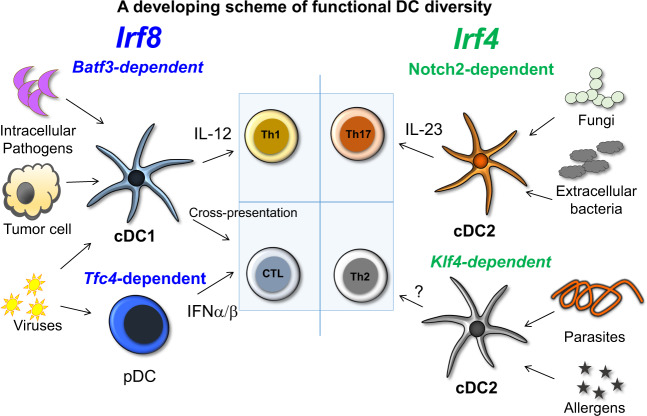
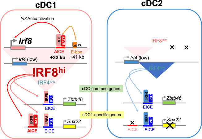
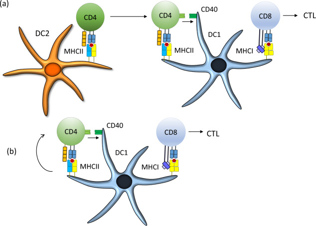
The clinical success of immune checkpoint therapy (ICT) has produced explosive growth in tumor immunology research because ICT was discovered through basic studies of immune regulation. Much of the current translational efforts are aimed at enhancing ICT by identifying therapeutic targets that synergize with CTLA4 or PD1/PD-L1 blockade and are solidly developed on the basis of currently accepted principles. Expanding these principles through continuous basic research may help broaden translational efforts. With this mindset, we focused this review on three threads of basic research directly relating to mechanisms underlying ICT. Specifically, this review covers three aspects of dendritic cell (DC) biology connected with antitumor immune responses but are not specifically oriented toward therapeutic use. First, we review recent advances in the development of the cDC1 subset of DCs, identifying important features distinguishing these cells from other types of DCs. Second, we review the antigen-processing pathway called cross-presentation, which was discovered in the mid-1970s and remains an enigma. This pathway serves an essential in vivo function unique to cDC1s and may be both a physiologic bottleneck and therapeutic target. Finally, we review the longstanding field of helper cells and the related area of DC licensing, in which CD4 T cells influence the strength or quality of CD8 T cell responses. Each topic is connected with ICT in some manner but is also a fundamental aspect of cell-mediated immunity directed toward intracellular pathogens.

Keywords: CD4 help; Dendritic cells; cross-presentation; transcription factors; tumor rejection.


Conflict of interest statement

The authors declare no competing interests.

Figures

Fig. 1
Fig. 1. Proposed scheme showing functional DC diversity.
The genetic basis of functional DC diversity is shown with IRF8-dependent types, cDC1s and pDCs, shown on the left. Both cDC1s and pDCs function in antiviral immunity, with pDCs contributing type I interferon and cDC1 priming CD8 T cells through cross-presentation of antigens. cDC1s are also active in some intracellular infections, such as in Toxoplasma gondii, where their IL-12 production in response to pathogens is key for host defense. cDC1s also prime CD8 T cells to recognize tumor cell antigens through cross-presentation. cDC2s can aid in protection from other pathogens, and evidence suggests that defense against some pathogens, such as Citrobacter rodentium, relies on a Notch2-dependent program, while defense against helminths relies on KLF4 action in cDC2s.
Fig. 2
Fig. 2. The basis for cDC1/cDC2 diversity lies in the activation of an AICE-dependent gene program.
In cDC1s, the Irf8 gene undergoes autoactivation through the action of an IRF8:BATF3 complex binding to an enhancer located +32 kb downstream of the Irf8 promoter. This autoactivation maintains high levels of IRF8 expression in the maturing cDC1 of a specific progenitor. In contrast, in cDC2s, the level of Irf8 is not maintained, and both IRF4 and IRF8 are expressed together but at much lower levels. This level of IRF4 and IRF8 expressed in cDC2s is sufficient to activate the EICE-dependent program that controls the expression of genes expressed in cDCs, both in cDC1s and cDC2s, such as Zbtb46. However, in cDC1s, the expression of IRF8 is much higher and sufficient to activate the program of genes that also require occupation of AICE cis-acting elements, such as the cDC1-specific gene Snx22. In Irf4 deficiency, a low level of IRF8 protein is sufficient to maintain the expression of some common cDC genes, but the induction of certain genes is inadequate for induction of some certain responses.
Fig. 3
Fig. 3. WDFY4 is a BEACH domain-containing protein required for cross-presentation by cDC1s in vivo and in vitro.
The WDFY4 protein shares similarity with other members of the BEACH domain-containing protein family. The PH (green) and BEACH (blue) domains comprise nearly one-third of a protein, lying in the carboxy-terminal region along with other domains, namely, WD40 and FYVE repeats (brown). The first approximately 2000 amino acids lack defined domains, although ongoing research suggests that this region contains at least one larger armadillo domain. The precise mechanism by which this protein plays a required role in cross-presentation is unknown despite high profile speculations.
Fig. 4
Fig. 4. Revised model of CD4 T cell licensing of cDC1s to induce CD8 T cell responses.
a In one model, CD4 T cells must be primed by cDC2s that have captured and presented tumor-derived antigens. These CD4 T cells migrate to the location of a cDC1 involved in priming CD8 T cells. b In a revised model, cDC1s serve as platforms for priming both CD4 T cells and CD8 T cells. The ligation of CD40 on the cDC1 surface during CD4 T cell priming is essential for strong licensing of cDC1s, and the lack of this ligation can lead to a failure in tumor rejection mediated by CD8 T cells. The mechanism downstream of CD40 signaling in cDC1s that leads to enhancement of CD8 responses is not currently known but may involve the induction of several costimulatory molecules, such as CD70 and 41BB, that augment the activation of CD8 T cells, as well as survival factors within the cDC1sthemselves.

References

    1. Ardavin C, Shortman K. Cell surface marker analysis of mouse thymic dendritic cells. Eur J Immunol. 1992;22:859–62. - PubMed
    1. Johansson-Lindbom B, Svensson M, Pabst O, Palmqvist C, Marquez G, Förster R, et al. Functional specialization of gut CD103(+) dendritic cells in the regulation of tissue-selective T cell homing. J Exp Med. 2005;202:1063–73. - PMC - PubMed
    1. Crozat K, Tamoutounour S, Vu Manh TP, Fossum E, Luche H, Ardouin L, et al. Cutting edge: expression of XCR1 defines mouse lymphoid-tissue resident and migratory dendritic cells of the CD8alpha+ type. J Immunol. 2011;187:4411–5. - PubMed
    1. Guilliams M, Ginhoux F, Jakubzick C, Naik SH, Onai N, Schraml BU, et al. Dendritic cells, monocytes and macrophages: a unified nomenclature based on ontogeny. Nat Rev Immunol. 2014;14:571–8. - PMC - PubMed
    1. Hildner K, Edelson BT, Purtha WE, Diamond M, Matsushita H, Kohyama M, et al. Batf3 deficiency reveals a critical role for CD8alpha+ dendritic cells in cytotoxic T cell immunity. Science. 2008;322:1097–1100. - PMC - PubMed