. 2021 Nov 4;81(21):4413-4424.e5.
doi: 10.1016/j.molcel.2021.08.006. Epub 2021 Sep 3.

SPT5 stabilization of promoter-proximal RNA polymerase II

Affiliations

SPT5 stabilization of promoter-proximal RNA polymerase II

Yuki Aoi et al. Mol Cell. .

Abstract

Based on in vitro studies, it has been demonstrated that the DSIF complex, composed of SPT4 and SPT5, regulates the elongation stage of transcription catalyzed by RNA polymerase II (RNA Pol II). The precise cellular function of SPT5 is not clear, because conventional gene depletion strategies for SPT5 result in loss of cellular viability. Using an acute inducible protein depletion strategy to circumvent this issue, we report that SPT5 loss triggers the ubiquitination and proteasomal degradation of the core RNA Pol II subunit RPB1, a process that we show to be evolutionarily conserved from yeast to human cells. RPB1 degradation requires the E3 ligase Cullin 3, the unfoldase VCP/p97, and a novel form of CDK9 kinase complex. Our study demonstrates that SPT5 stabilizes RNA Pol II specifically at promoter-proximal regions, permitting RNA Pol II release from promoters into gene bodies and providing mechanistic insight into the cellular function of SPT5 in safeguarding accurate gene expression.

Keywords: CDK9; Cullin 3; NELF; RNA polymerase II; SPT5; VCP; auxin-inducible degron; degradation; elongation; transcription.


Conflict of interest statement

Declaration of interests The authors declare no competing interests.

Figures

Figure 1.
Figure 1.. Rapid SPT5 depletion leads to RPB1 degradation.
(A) Schematic of auxin-inducible degradation of SPT5-AID protein. (B) Western blots of the indicated proteins in whole-cell lysates of SPT5-AID or parental DLD-1 cells. Cells were treated with 500 µM auxin for the indicated time. SPT5-AID was depleted within 2 h of auxin treatment, at which time the RPB1 loss was observed. HSP90 serves as a loading control. A triangle indicates increasing amounts of lysates. RPB1 NTD antibody D8L4Y was used to detect total RPB1 levels. (C, D) Western blots in NELF-C-AID (C) or PAF1-AID (D) cells treated with auxin as in (B). RPB1 levels were stable upon depletion of NELF or PAF1. RPB1 NTD antibody D8L4Y was used to detect total RPB1 levels. (E) Western blots in SPT5-AID cells treated with translation inhibitor cycloheximide (CHX) for 4 h, followed by treatment with 500 µM auxin for 3 h. RPB1 loss was still observed when protein synthesis was blocked, indicating RPB1 degradation upon SPT5 depletion. RPB1 NTD antibody D8L4Y was used to detect total RPB1 levels. See also Figure S1.
Figure 2.
Figure 2.. CUL3, VCP and PSMD12 are recruited to Pol II upon SPT5 depletion.
(A) MA plot showing mass spectrometric data of Pol II immunoprecipitation from 3 independent experiments. Proteins with > 2.5-fold change and p-value < 0.05 are shown in orange. Pol II subunits are shown in dark gray. See also Figure S2 and Table S1. (B) Log2 fold change of Pol II-associated complexes. Each dot indicates a subunit of each complex that was detected in our proteomics data. Dashed lines indicate 2.5-fold change.
Figure 3.
Figure 3.. E3 ligase CUL3, but not NEDD4, is required for RPB1 degradation after SPT5 depletion.
(A) Western blots of the indicated proteins in SPT5-AID7 whole-cell lysates. Cells were treated with shRNA against CUL3 or NEDD4, followed by treatment with 500 µM auxin for 3 h. RPB1 NTD antibody D8L4Y was used to detect total RPB1 levels. (B) Representative track showing Pol II ChIP-seq signal in SPT5-AID7 cells. Cells were treated as in (A). RPB1 NTD antibody D8L4Y was used to detect total RPB1 signal. (C) Meta plots of Pol II ChIP-seq in SPT5-AID7 cells treated as in (A). Signal is centered on promoter-proximal pause site. N = 6,524. (D) Heat map of ChIP-seq signal for Pol II and CUL3. Rows are sorted by Pol II occupancy levels. SPT5-AID cells were treated with or without 500 µM auxin for 1.5 h. Note that auxin treatment for 1.5 h leads to a limited degradation of RPB1. N = 6,524. See also Figure S3.
Figure 4.
Figure 4.. Unfoldase VCP is required for RPB1 degradation induced by SPT5 depletion in human and yeast.
(A) VCP knockdown rescued RPB1 degradation. SPT5-AID cells were treated with the indicated shRNA, followed by treatment with 500 µM auxin for the indicated time. NT, non-targeting. Actin serves as a loading control. RPB1 NTD antibody D8L4Y was used to detect total RPB1 levels. (B) Pharmacological inhibition of VCP rescued RPB1 degradation. SPT5-AID cells were treated with 2.5 µM VCP inhibitor NMS-873 (VCPi) for 4 h, followed by treatment with 500 µM auxin for the indicated time. Actin serves as a loading control. RPB1 NTD antibody D8L4Y was used to detect total RPB1 levels. (C) Inhibition of transcription initiation led to VCP-independent RPB1 degradation. SPT5-AID cells were treated with 2.5 µM NMS-873 (VCPi) for 5 h, followed by treatment with a range of dosage of initiation inhibitor triptolide (TPL) for 3 h. As a control, cells were treated with 500 µM auxin instead of TPL. RPB1 NTD antibody D8L4Y was used to detect total RPB1 levels. See also Figure S4.
Figure 5.
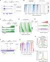
Figure 5.. SPT5 depletion results in destabilization of Pol II during early elongation.
(A) Representative genome browser track example showing ChIP-seq signal for Pol II (blue) and SPT5 (purple) in SPT5-AID cells. Cells were treated with 2.5 µM NMS-873 (VCPi) for 4 h, followed by treatment with 500 µM auxin for 3 h. RPB1 NTD antibody D8L4Y was used to detect total RPB1 signal. (B) Heat map of ChIP-seq for Pol II and SPT5. SPT5-AID cells were treated as in (A). Rows are sorted by Pol II occupancy levels in the vehicle (untreated) sample. N = 6,524. (C) Pulse-chase nascent RNA labeling assay. SPT5-AID cells were treated with 2.5 µM NMS-873 (VCPi) for 4 h, then 500 µM auxin for 3 h. After that, cells were treated with 250 nM P-TEFb inhibitor NVP-2 for 1 h, followed by nascent RNA labeling with 500 µM 4sU for 15 min. Representative tracks of TT-seq signal (labeled nascent RNA) is shown. Inhibition of P-TEFb blocks pause escape but not elongation of Pol II that has already escaped (Jonkers et al., 2014). Horizontal black lines along tracks indicate the distance that Pol II travels for 1 h. (D) Heat maps of TT-seq signal in SPT5-AID cells treated as in (C). Rows are sorted by gene length. Curved lines in heat maps indicate transcription end site (TES). N = 2,386 genes with length of ≥50 kb. (E) Rain cloud plot showing single-gene level estimates of Pol II elongation rate. N = 69 genes with length of > 240 kb and >100 rpm. **** indicates p < 3 × 10⁻⁷ and ns indicates “not significant” (p = 0.058) in Mann Whitney U test. (F) Representative track showing PRO-seq signal in SPT5-AID cells at promoter-proximal regions. Cells were treated as in (A). (G) Heat map of PRO-seq signal. The left panel shows PRO-seq signal in the untreated condition (vehicle), and the right 2 panels show log2 fold change relative to vehicle. N = 6,524. (H) Meta plot of PRO-seq signals in SPT5-AID cells treated as in (A). Signal is centered on the +1 nucleosome dyad. Light gray box indicates the position of the +1 nucleosome (dyad ± 75 bp) as determined by MNase-seq. N = 1,843. (I) Meta profile of NELF ChIP-seq signal at promoter-proximal regions. SPT5-AID cells were treated as in (A). See also Figure S5.
Figure 6.
Figure 6.. P-TEFb is required for RPB1 degradation induced by SPT5 loss.
(A) Representative track showing Pol II ChIP-seq signal in SPT5-AID cells. Cells were treated with 250 nM P-TEFb inhibitor NVP2 or CDK9 degrader THAL-SNS-032 (dCDK9) for 2 h, followed by treatment with 500 µM auxin for 3 h. RPB1 NTD antibody D8L4Y was used to detect total RPB1 signal. (B) Heat map of Pol II ChIP-seq in SPT5-AID cells treated as in (A). Rows are sorted by Pol II occupancy levels. N = 6,524. (C) Meta plot of Pol II ChIP-seq in SPT5-AID cells treated as in (A). Signal is centered on promoter-proximal pause site. N = 6,524. (D) Western blots of the indicated proteins in SPT5-AID whole-cell lysates. Cells were pretreated with 250 nM NVP-2, 250 nM dBET6, 20 µM KL1 or KL2 for 4 h, followed by 500 µM auxin treatment for 4 h. RPB1 NTD antibody D8L4Y was used to detect total RPB1 levels. See also Figure S6.
Figure 7.
Figure 7.. Model: SPT5 stabilizes promoter-proximal Pol II.
(A) SPT5 functions in stabilizing Pol II during the early elongation stage. With SPT5 protein (upper panel), once Pol II is initiated and escapes from the promoter, Pol II undergoes productive elongation until termination. Without SPT5 (lower panel), early elongating Pol II undergoes RPB1 degradation that is mediated by the CUL3 ubiquitin ligase, the VCP unfoldase, and a novel form of P-TEFb component CDK9. Late elongating Pol II without SPT5 can persist in transcription at a slower speed. (B) A failure in Pol II passage on the +1 nucleosome leads to 2 distinct pathways to eliminate Pol II from chromatin.

References

    1. Anindya R, Aygün O, and Svejstrup JQ (2007). Damage-induced ubiquitylation of human RNA polymerase II by the ubiquitin ligase Nedd4, but not Cockayne syndrome proteins or BRCA1. Molecular Cell 28, 386–397. - PubMed
    1. Aoi Y, Smith ER, Shah AP, Rendleman EJ, Marshall SA, Woodfin AR, Chen FX, Shiekhattar R, and Shilatifard A. (2020). NELF Regulates a Promoter-Proximal Step Distinct from RNA Pol II Pause-Release. Molecular Cell 78, 261–274.e265. - PMC - PubMed
    1. Banerjee S, Bartesaghi A, Merk A, Rao P, Bulfer SL, Yan Y, Green N, Mroczkowski B, Neitz RJ, Wipf P., et al. (2016). 2.3 Å resolution cryo-EM structure of human p97 and mechanism of allosteric inhibition. Science 351, 871–875. - PMC - PubMed
    1. Baumli S, Hole AJ, Wang L-Z, Noble MEM, and Endicott JA (2012). The CDK9 tail determines the reaction pathway of positive transcription elongation factor b. Structure 20, 1788–1795. - PMC - PubMed
    1. Booth GT, Wang IX, Wang IX, Cheung VG, Cheung VG, and Lis JT (2016). Divergence of a conserved elongation factor and transcription regulation in budding and fission yeast. Genome Res. 26, 799–811. - PMC - PubMed

Publication types

MeSH terms