Review
. 2022 Mar:231:107977.
doi: 10.1016/j.pharmthera.2021.107977. Epub 2021 Sep 1.

G protein-coupled receptor-effector macromolecular membrane assemblies (GEMMAs)

Affiliations
Review

G protein-coupled receptor-effector macromolecular membrane assemblies (GEMMAs)

Sergi Ferré et al. Pharmacol Ther. 2022 Mar.

Abstract

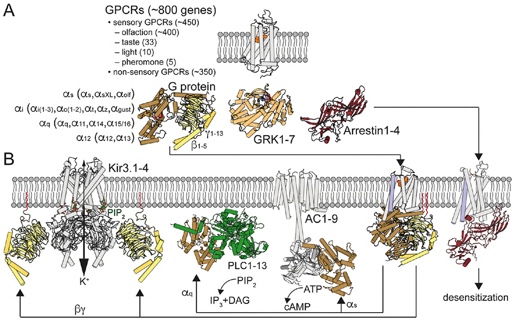
G protein-coupled receptors (GPCRs) are the largest group of receptors involved in cellular signaling across the plasma membrane and a major class of drug targets. The canonical model for GPCR signaling involves three components - the GPCR, a heterotrimeric G protein and a proximal plasma membrane effector - that have been generally thought to be freely mobile molecules able to interact by 'collision coupling'. Here, we synthesize evidence that supports the existence of GPCR-effector macromolecular membrane assemblies (GEMMAs) comprised of specific GPCRs, G proteins, plasma membrane effector molecules and other associated transmembrane proteins that are pre-assembled prior to receptor activation by agonists, which then leads to subsequent rearrangement of the GEMMA components. The GEMMA concept offers an alternative and complementary model to the canonical collision-coupling model, allowing more efficient interactions between specific signaling components, as well as the integration of the concept of GPCR oligomerization as well as GPCR interactions with orphan receptors, truncated GPCRs and other membrane-localized GPCR-associated proteins. Collision-coupling and pre-assembled mechanisms are not exclusive and likely both operate in the cell, providing a spectrum of signaling modalities which explains the differential properties of a multitude of GPCRs in their different cellular environments. Here, we explore the unique pharmacological characteristics of individual GEMMAs, which could provide new opportunities to therapeutically modulate GPCR signaling.

Keywords: G protein subnits; G protein-coupled receptors; GPCR allosterism; GPCR oligomerization; Plasma membrane effector.


Conflict of interest statement

Declaration of Competing Interest J.G.-M. has a sponsored research contract with Neuristic Pharma. The remaining authors declare that there are no conflicts of interest.

Figures

Fig. 1.
Fig. 1.. G protein-coupled receptor signaling.
(A) GPCRs are illustrated with the structure of retinal-bound rhodopsin in white (Palczewski et al., 2000). In humans, there are 16 Gα subunits (classified as Gαs, Gαi, Gαq and Gα12) 5 Gβ and 12 Gγ (Gγ1-13, but no Gγ6) subunits. Active GPCRs interact with Gαβγ proteins, illustrated with the structure of GDP-bound Gαi1β1γ2 in brown/yellow (Wall et al., 1995), G protein-coupled receptor kinases (GRKs), with 7 mammalian isoforms, illustrated with the structure of ATP-bound GRK1 in light orange (Singh, Wang, Maeda et al., 2008), and arrestins, with 4 isoforms, illustrated with the structure of arrestin1 in red (Granzin et al., 1998). (B) Agonist binding triggers the movement of TM6 (in blue), opening an intracellular cavity for G protein arrangement, illustrated with the crystal structure of the agonist-β2AR-Gs complex (Rasmussen et al., 2011). Gαq activates PLC, with 13 mammalian isozymes, illustrated with the crystal structure of PLC-β3 bound to Gαq (Waldo et al., 2010), which catalyzes the hydrolysis of PIP2 to the Ca2+-mobilizing second messenger inositol 1,4,5-trisphosphate (IP3) and the PKC-activating second messenger diacylglycerol (DAG). Gαs stimulates and Gαi inhibits the activity of AC, with 9 mammalian isoforms, illustrated with the cryo-EM structure of AC9 bound to Gαs (Qi et al., 2019). Gβγ subunits increase the activity of Kir3 channels, illustrated with the crystal structure of the Kir3.2-Gβγ (Whorton and MacKinnon, 2011). Desensitization is mediated by GRK-mediated GPCR phosphorylation and subsequent binding of arrestin, illustrated with the cryo-EM structure of neurotensin NT1 receptor in complex with arrestin2 (Huang et al., 2020). Gγ isoprenylated group is shown in red.
Fig. 2.
Fig. 2.. Molecular structures of GPCR homomers.
(A) Cryo-EM structures of the full length mGlu5R in the apo inactive/open and active/closed onformations (Koehl et al., 2019). Class C GPCRs are obligate cysteine-linked homomers and each protomer is formed by a large extracellular ‘Venus fly-trap’ domain that binds orthosteric ligands and promotes an inter-protomer disulfide bridge formed via a conserved cysteine, a cysteine rich domain that connects these two domains and a seven TM domain (7TM). The helix interface between protomers of the Venus fly-trap domains is represented by cylinders. Activation leads to compacting of the mGlu5R homomer, bringing the cysteine rich domains into proximity and enabling the 7TM domains to approximate to initiate signaling. The inactive state shows a TM5-TM5 orientation but with substantial separation between the 7TM domains. Activation leads to a 20° rotation of each 7TM domain, resulting in a close contact TM6-TM6 homodimeric interface. Both protomers are colored in light blue and gray. (B) CXC4R structure (Wu et al., 2010) reveals a homomer with an interface including TM5 and TM6. (C) MOR structure (Manglik et al., 2012) shows receptor protomers associated in pairs through two different interfaces. One interface is via TM1, TM2 and helix-8 (Hx8) (blue/white and light/dark green protomers) and the other interface comprises TM5 and TM6 (white/green protomers). (D) β1AR structure (Huang, Chen, Zhang, and Huang, 2013)15 also displays two homomeric interfaces. One interface also involves TM1, TM2 and Hx8 (blue/white and light/dark green protomers), as with MOR. In contrast, the other interface engages residues from TM4, TM5 and intracellular loop 2 (ICL2) white/green protomers).
Fig. 3.
Fig. 3.. Scaffolding of GPCRs, ligand-gated ion channels and PM-effectors mediated by PDZ proteins and AKAPs.
(A) Complexes of ionotropic glutamate AMPAR group I metabotropic receptors (mGlu1R or mGlu5R) established by a chain of interactions of the scaffold proteins PSD-95, GKAP, Shank and Homer. Homer directly interacts with the CT of mGlu1R or mGl5R, while AMPAR indirectly binds to PSD-95 by oligomerization with the TM AMPAR regulatory protein stargazing (Str). (B) Complexes of β2AR, AC and L-type voltage-dependent Cav1.2 Ca2+ channels mediated by AKAP5. (C) Complexes of β2AR, AC and AMPAR mediated by AKAP5 and PSD-95. By bringing together β2AR, AC and PKA, AKAP5-mediated complexes provide the frame for an efficient β2AR-dependent, PKA-mediated phosphorylation of the α1.2 subunit of Cav1.2 channels (B) and the GluA1 subunit of AMPAR (C).
Fig. 4.
Fig. 4.. Allosterism in GPCR heteromers.
(A) Type I allosterism, in which a ligand (red spheres) binding to one protomer (green cylinders) in the GPCR heteromer can modify the affinity (ligand binding depicted by the red arrow) or the efficacy (receptor activation depicted by the wide orange arrow) of another ligand (orange spheres) binding to the partner receptor (gray cylinders) via their TM helices (see Fig. 5). (B) Type II allosterism, or ligand-independent allosterism, in which the affinity or efficacy of a ligand (orange spheres) for a GPCR (gray cylinders) can be modified just by heteromerization with a molecularly different GPCR (green cylinders). (C) Lateral view (left) and extracellular view (right) of a computer model of a GEMMA including two heterotetramers composed of two different GPCR homomers (one represented by white cylinders and grey surfaces and the other represented by green cylinders and surfaces), AC (light blue cylinders; based on the cryo-EM structure of AC9; Qi et al, 2019), Gs (brown for Gαs and yellow for Gβγ) and Gi (red for Gαi and yellow for Gβγ) (based on Navarro et al., 2018). This type of GEMMA can explain the ability of a Gi-coupled GPCR to counteract AC activation mediated by a Gs-coupled GPCR (type III allosterism). The proposed contact between the CT domain of the receptor and the Gβ subunit is shown by a color line (Tsai et al. 2019). To facilitate visualization of all protomers of the GPCR oligomers, one of the protomers is represented by cylinders and the other protomers are represented as color surfaces (with different shades of the same color for the same GPCR). Both positions of the Gαs and Gαi subunits, in their Gβγ-associated and receptor-bound and state (without surface) and in their Gβγ-dissociated and AC-bound state (with surface), are shown. The agonist-induced dissociation of Gαs and Gαi from their respective Gβγ takes place within the framework of the GEMMA..
Fig. 5.
Fig. 5.. Mechanisms of type I allosterism in GPCR oligomers heteromers.
(A) Cross-section through a prototypical class A GPCR, highlighting the agonist (white sticks), the amino acids of the orthosteric site (delineated by a white line) involved in ligand binding (orange), the conserved PIF motif (yellow), and the NPxxY and DRY motifs and Y, (yellow) that transmit the signal from the PIF motif (transmission switch) to the G protein site (Weis and Kobilka, 2018). (B) Position of these amino acids involved in ligand binding (orange spheres) and signal transmission (yellow) in models of GPCR dimers via TM5 and TM6 (top) and TM4 and TM5 and intracellular loop 2 (bottom). Clearly, a ligand-bound (type I allosterism) or ligand-free (type II allosterism) or orphan GPCR (green cylinders) can modify the affinity (orange spheres) or efficacy (yellow spheres) of the partner receptor (gray) via the protein-protein interface (blue mesh). (C) GPCRs are dynamic proteins that adopt, in a ligand-free form, a number of conformations (shades in gray) that not only involve the extracellular and intracellular sites (Weis and Kobilka, 2018), but also a potential TM oligomeric interface. An inverse agonist (red polygon) binding to one of the protomers of a GPCR heteromer triggers a high surface complementarity between TM5 and TM6 of the two molecularly different protomers, via the four-helix bundle (dashed red circle) (Manglik et al., 2012), which blocks the opening of the intracellular cavity for G protein binding at the other protomer (cross-antagonism) (Viñals et al., 2015). An agonist (green polygon) binding to one of the protomers triggers the outward movement of TM 6 (see arrows), opening the intracellular cavity for G protein binding (Rasmussen et al., 2011), whereas the TM5 and TM6 heteromeric interface impedes simultaneous agonist-induced movement of both TM6 (negative crosstalk) due to a steric clash (red cross) (Guinart et al., 2020).
Fig. 6.
Fig. 6.. The A2AR-D2R heterotetramer-AC5 and mGlu2R-5-HT2AR-PLC-Kir3 GEMMAs.
(A) The A2AR-D2R heterotetramer-AC5 GEMMA, constituted by A2AR homomers coupled to Gs, D2R homomers coupled to Gi and AC5, can be considered as a prototype of GEMMA that integrates canonical antagonistic Gs-Gi-AC interactions. (B) The mGlu2R-5-HT2AR-PLC-Kir3 GEMMA, constituted by mGlu2R homomers coupled to Gi, 5-HT2AR homomers coupled to Gq, PLC and Kir3, can be considered as a prototype of GEMMA that integrates canonical antagonistic Gi-Gq-Kir3 interactions.
Fig, 7.
Fig, 7.. Oligomerization of GPCRs with orphan GPCRs, truncated GPCRs and RAMPs.
(A) Homomers and heteromers of MT1R, MT2R and GPR50. MT2R preferentially exists as a heteromeric complex with MT1R and MT1R-MT2R heteromerization drives a change in the preference of G protein subtype coupling, from Gi to Gq. The long CT of GPR50 significantly alters MT1R-Gi protein signaling in the MT1R-GPR50 heteromer (see text). (B) GHS1aR forms homomers and oligomerizes with its non-functional truncated isoform GHS1bR to form homomers and heteromers. One of the properties of the GHS1aR-GHS1bR oligomers is the facilitation of an additional interaction with the D1R, leading to a change in the preference of G protein subtype coupling of GHS1aR, from Gq to Gs. (C) The single TM domain proteins RAMP1, RAMP2 and RAMP3 associate with CLR and form the Gs protein-coupled receptors CGRP, AM1R and AM2R, respectively, determining the ligands potentially binding to CLR.

References

    1. Ahmed SN, Brown DA, & London E (1997). On the origin of sphingolipid/cholesterol-rich detergent-insoluble cell membranes: physiological concentrations of cholesterol and sphingolipid induce formation of a detergent-insoluble, liquid-ordered lipid phase in model membranes. Biochemistry, 36(36), 10944–10953. - PubMed
    1. Andressen KW, Ulsund AH, Krobert KA, Lohse MJ, Bünemann M, & Levy FO (2018). Related GPCRs couple differently to Gs: preassociation between G protein and 5-HT7 serotonin receptor reveals movement of Gαs upon receptor activation. FASEB Journal, 32(2), 1059–1069. - PubMed
    1. Ayoub MA, Couturier C, Lucas-Meunier E, Angers S, Fossier P, Bouvier M, et al. (2002). Monitoring of ligand-independent dimerization and ligand-induced conformational changes of melatonin receptors in living cells by bioluminescence resonance energy transfer. The Journal of Biological Chemistry, 277(24), 21522–21528. - PubMed
    1. Ayoub MA, Levoye A, Delagrange P, & Jockers R (2004). Preferential formation of MT1/MT2 melatonin receptor heterodimers with distinct ligand interaction properties compared with MT2 homodimers. Molecular Pharmacology, 66(2), 312–321. - PubMed
    1. Ayoub MA, Trinquet E, Pfleger KD, & Pin JP (2010). Differential association modes of the thrombin receptor PAR1 with Galphai1, Galpha12, and beta-arrestin 1. FASEB Journal, 24(9), 3522–3535. - PubMed

Publication types

MeSH terms

Substances