

Surfactants influence polymer nanoparticle fate within the brain
- PMID: 34481289
- PMCID: PMC8478896
- DOI: 10.1016/j.biomaterials.2021.121086
Surfactants influence polymer nanoparticle fate within the brain
Abstract
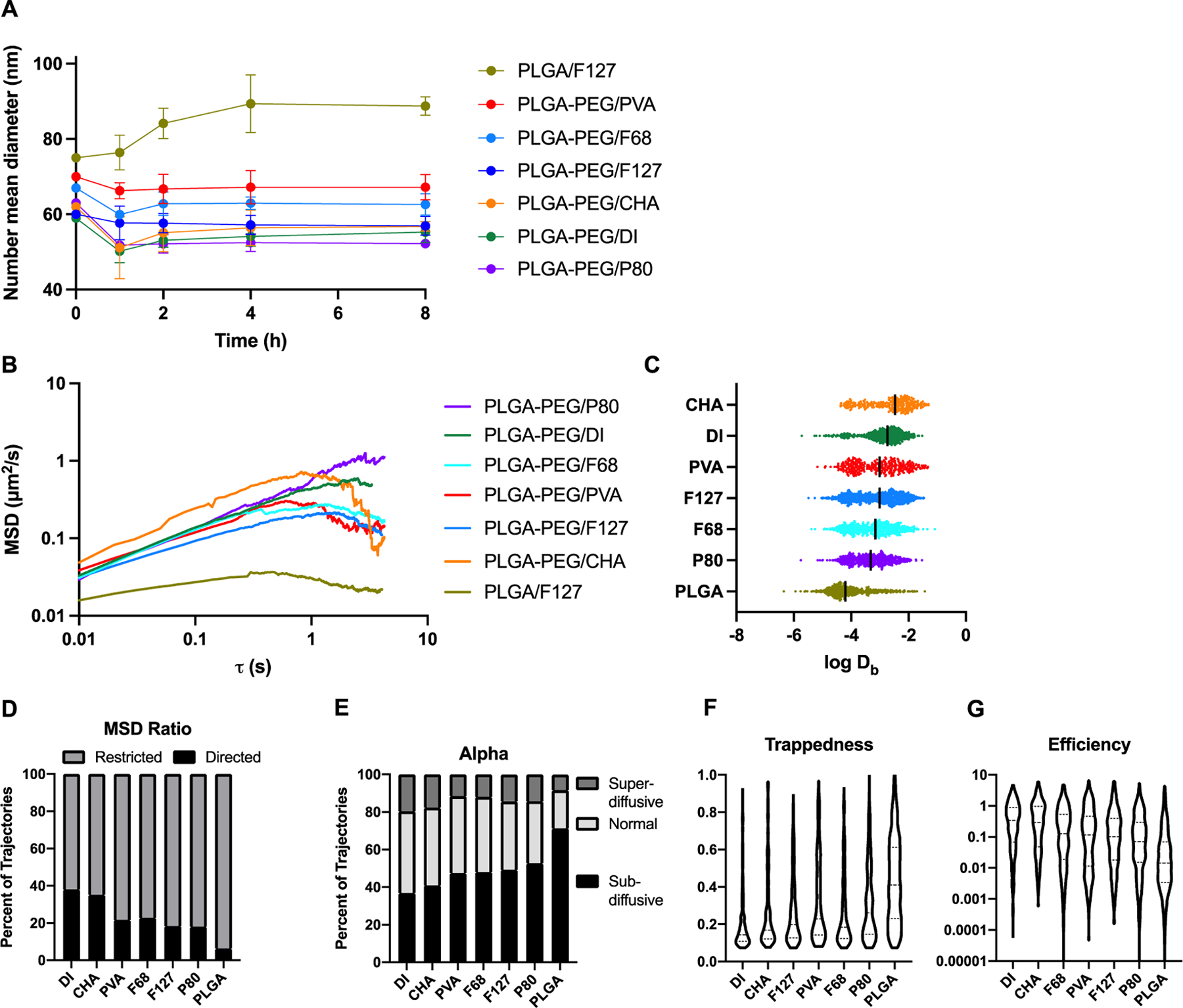
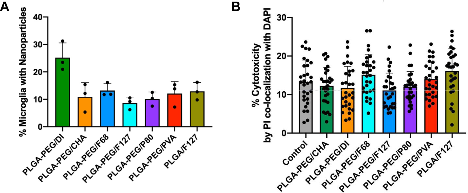
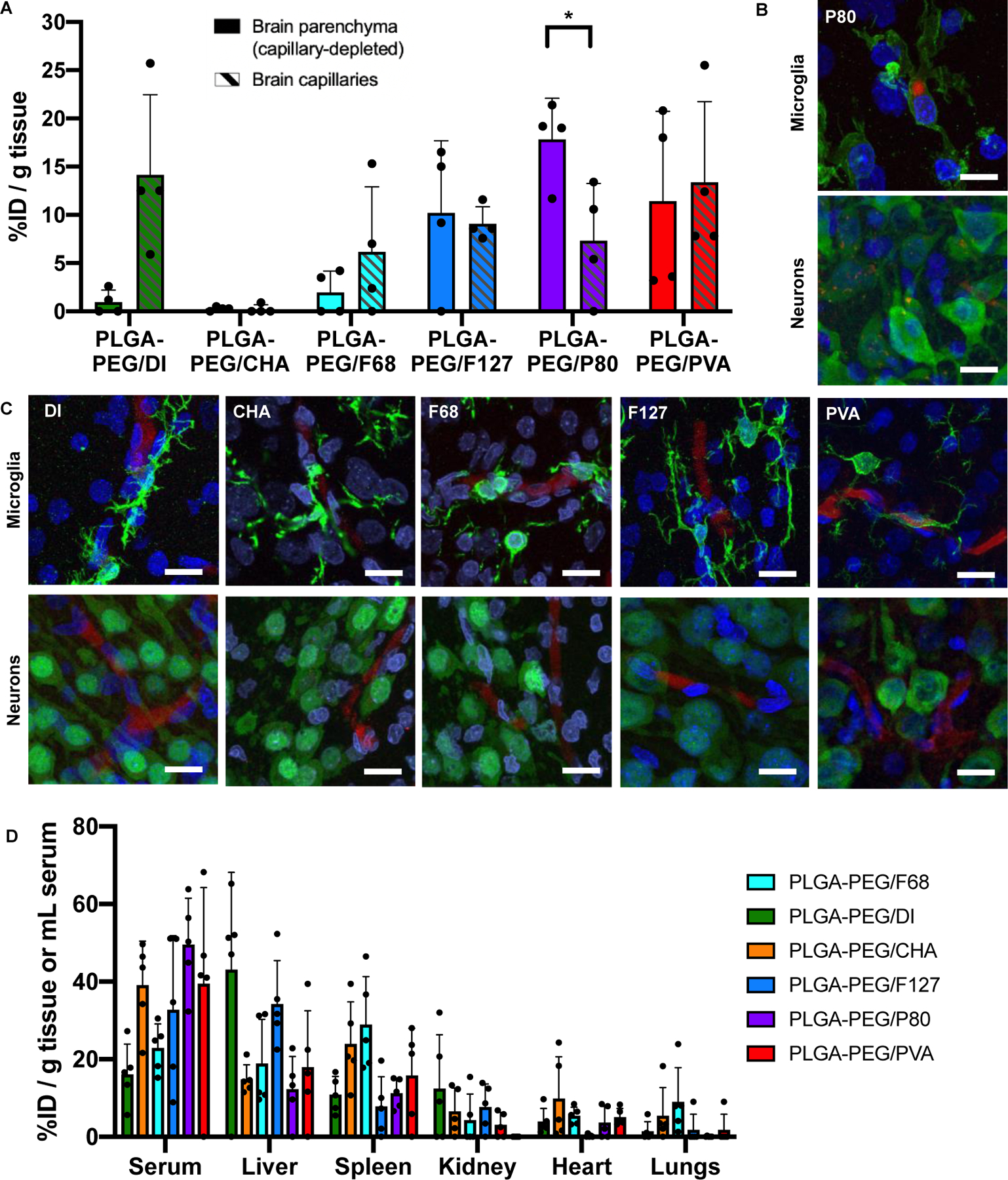
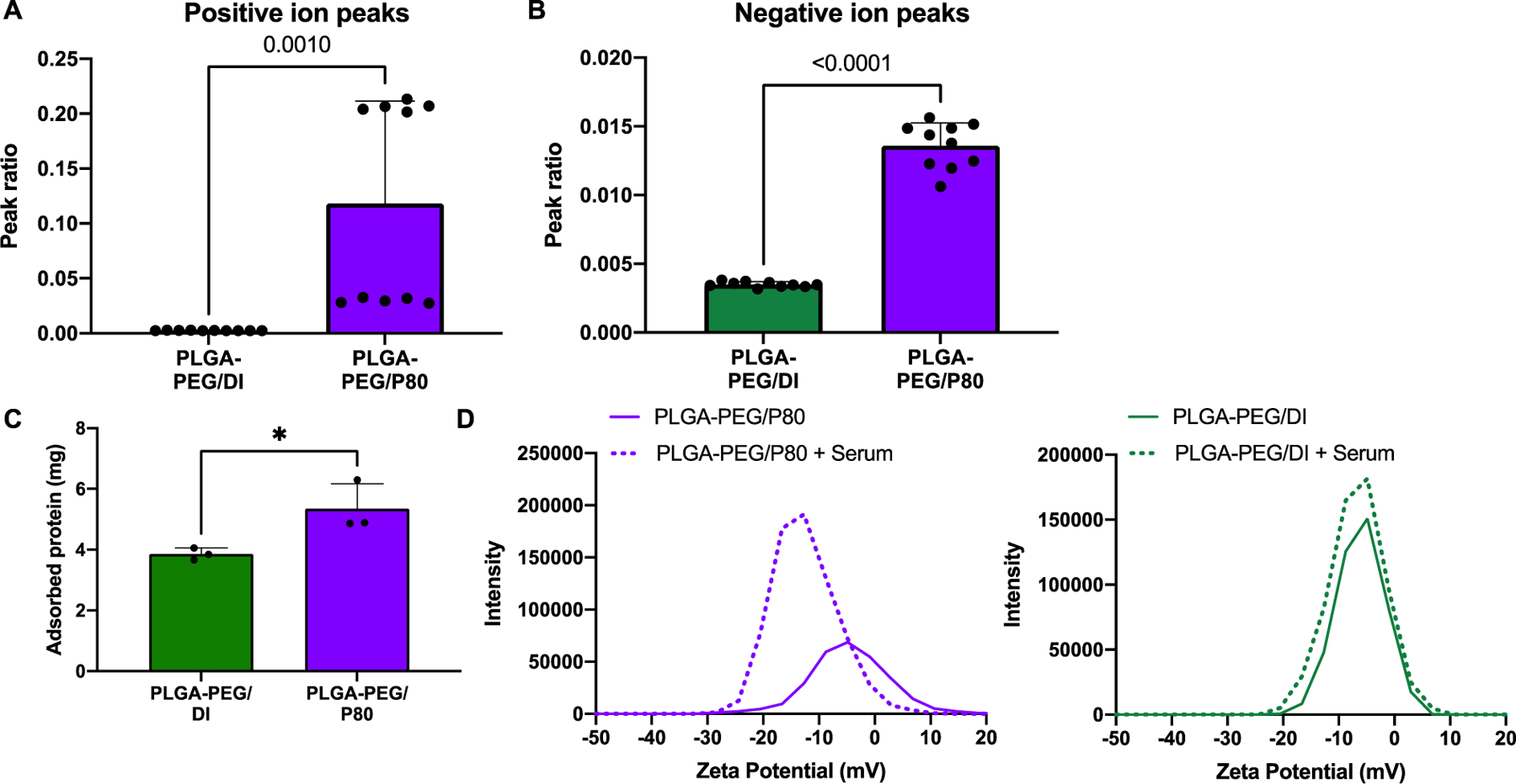
Drug delivery to the brain is limited by poor penetration of pharmaceutical agents across the blood-brain barrier (BBB), within the brain parenchyma, and into specific cells of interest. Nanotechnology can overcome these barriers, but its ability to do so is dependent on nanoparticle physicochemical properties including surface chemistry. Surface chemistry can be determined by a number of factors, including by the presence of stabilizing surfactant molecules introduced during the formulation process. Nanoparticles coated with poloxamer 188 (F68), poloxamer 407 (F127), and polysorbate 80 (P80) have demonstrated uptake in BBB endothelial cells and enhanced accumulation within the brain. However, the impact of surfactants on nanoparticle fate, and specifically on brain extracellular diffusion or intracellular targeting, must be better understood to design nanotherapeutics to efficiently overcome drug delivery barriers in the brain. Here, we evaluated the effect of the biocompatible and commonly used surfactants cholic acid (CHA), F68, F127, P80, and poly (vinyl alcohol) (PVA) on poly (lactic-co-glycolic acid)-poly (ethylene glycol) (PLGA-PEG) nanoparticle transport to and within the brain. The inclusion of these surfactant molecules decreases diffusive ability through brain tissue, reflecting the surfactant's role in encouraging cellular interaction at short length and time scales. After in vivo administration, PLGA-PEG/P80 nanoparticles demonstrated enhanced penetration across the BBB and subsequent internalization within neurons and microglia. Surfactants incorporated into the formulation of PLGA-PEG nanoparticles therefore represent an important design parameter for controlling nanoparticle fate within the brain.
Keywords: Blood-brain barrier; Brain drug delivery; Cellular uptake; Diffusion; Polymeric nanoparticles; Surfactant.
Copyright © 2021 Elsevier Ltd. All rights reserved.
Conflict of interest statement
Conflict of Interest Disclosure
The authors declare no competing financial interest.
Declaration of interests
The authors declare that they have no known competing financial interests or personal relationships that could have appeared to influence the work reported in this paper.
Figures
References
-
- Wong HL; Wu XY; Bendayan R, Nanotechnological advances for the delivery of CNS therapeutics. Adv Drug Deliv Rev 2012, 64 (7), 686–700. - PubMed
-
- Tosi G; Costantino L; Ruozi B; Forni F; Vandelli MA, Polymeric nanoparticles for the drug delivery to the central nervous system. Expert Opin Drug Deliv 2008, 5 (2), 155–74. - PubMed
-
- Liu Z; Gao X; Kang T; Jiang M; Miao D; Gu G; Hu Q; Song Q; Yao L; Tu Y; Chen H; Jiang X; Chen J, B6 peptide-modified PEG-PLA nanoparticles for enhanced brain delivery of neuroprotective peptide. Bioconjug Chem 2013, 24 (6), 997–1007. - PubMed
-
- Joseph A; Wood T; Chen CC; Corry K; Snyder JM; Juul SE; Parikh P; Nance E, Curcumin-loaded polymeric nanoparticles for neuro-protection in neonatal rats with hypoxic-ischemic encephalopathy. Nano Res 2018, 11 (10), 5670–5688.
Publication types
MeSH terms
Substances
Grants and funding
LinkOut - more resources
Full Text Sources
Miscellaneous
