Clinical Trial
. 2022 Feb 24;139(8):1208-1221.
doi: 10.1182/blood.2021011354.

Identification and prioritization of myeloid malignancy germline variants in a large cohort of adult patients with AML

Affiliations
Clinical Trial

Identification and prioritization of myeloid malignancy germline variants in a large cohort of adult patients with AML

Fei Yang et al. Blood. .

Abstract

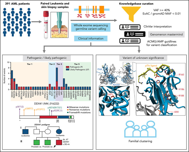
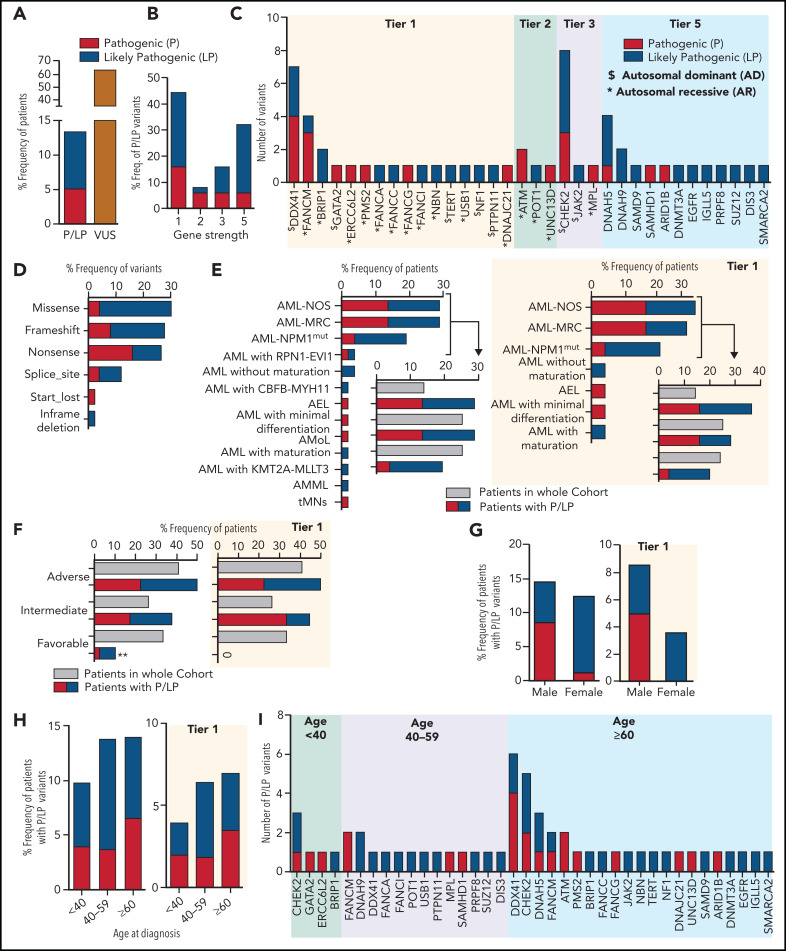
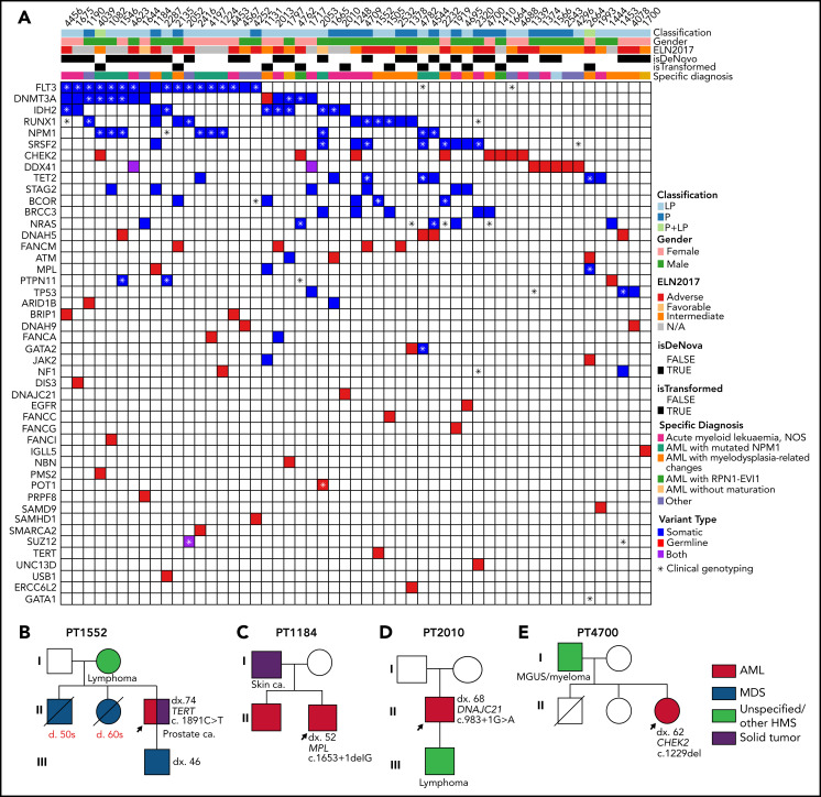
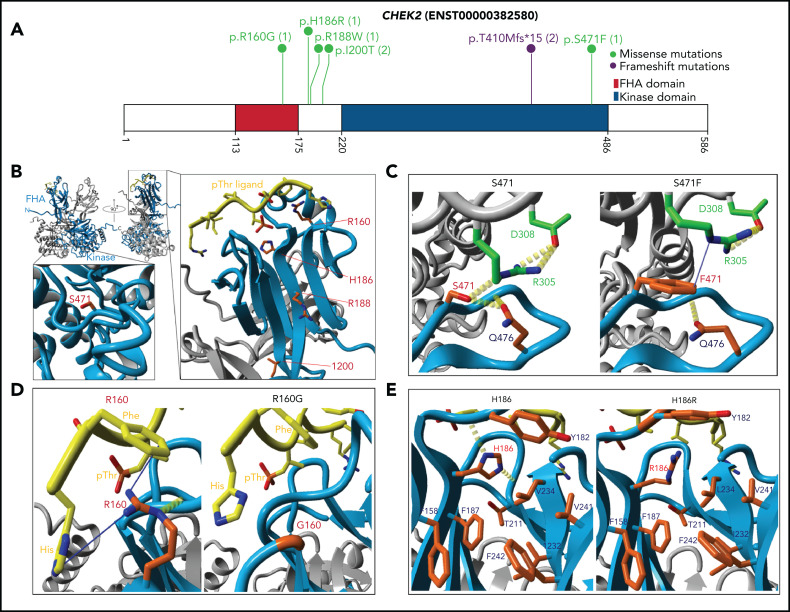
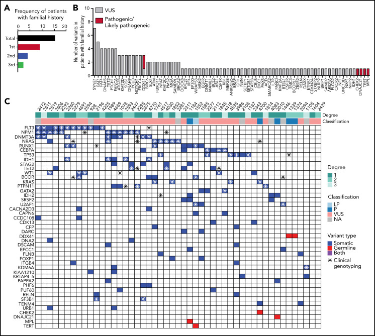
Inherited predisposition to myeloid malignancies is more common than previously appreciated. We analyzed the whole-exome sequencing data of paired leukemia and skin biopsy samples from 391 adult patients from the Beat AML 1.0 consortium. Using the 2015 American College of Medical Genetics and Genomics (ACMG) guidelines for variant interpretation, we curated 1547 unique variants from 228 genes. The pathogenic/likely pathogenic (P/LP) germline variants were identified in 53 acute myeloid leukemia (AML) patients (13.6%) in 34 genes, including 6.39% (25/391) of patients harboring P/LP variants in genes considered clinically actionable (tier 1). 41.5% of the 53 patients with P/LP variants were in genes associated with the DNA damage response. The most frequently mutated genes were CHEK2 (8 patients) and DDX41 (7 patients). Pathogenic germline variants were also found in new candidate genes (DNAH5, DNAH9, DNMT3A, and SUZ12). No strong correlation was found between the germline mutational rate and age of AML onset. Among 49 patients who have a reported history of at least one family member affected with hematological malignancies, 6 patients harbored known P/LP germline variants and the remaining patients had at least one variant of uncertain significance, suggesting a need for further functional validation studies. Using CHEK2 as an example, we show that three-dimensional protein modeling can be one of the effective methodologies to prioritize variants of unknown significance for functional studies. Further, we evaluated an in silico approach that applies ACMG curation in an automated manner using the tool for assessment and (TAPES) prioritization in exome studies, which can minimize manual curation time for variants. Overall, our findings suggest a need to comprehensively understand the predisposition potential of many germline variants in order to enable closer monitoring for disease management and treatment interventions for affected patients and families.


Figures

None
Graphical abstract
Figure 1.
Figure 1.
The pathogenic and likely pathogenic germline variants identified in 391 AML patients. (A) The percentage frequency of patients with pathogenic and likely pathogenic (P/LP) and variants of uncertain significance (VUS) in 391 patients. For patients 4039 and 2664, each has 2 germline variants, 1 in each P and LP category and thus counted once only in P category. (B) The percentage frequency of P and LP variants associated with gene strength for 50 unique variants. To define gene strength, 291 genes were selected for curation based on a comprehensive literature review. The strength of the genes is assigned from 1 to 5, depending on the evidence supporting their association with cancer predisposition to either hematological malignancies or other types of cancer, inherited hematological disorders that may predispose to hematological neoplasms, or reported as a somatic variant contributing to the pathogenesis of hematological malignancies. (C) Number of pathogenic and likely pathogenic germline variants in 34 genes for 56 variants in 53 patients, categorized by the gene strength. Variants in genes with “$” were associated with autosomal dominant conditions and those with asterisks were associated with AR syndromes. (D) Percentage frequency of variants classified by type of variants for 50 unique P/LP variants. (E-F) Association of P and LP variants with disease specific subgroups (E) or 2017 ELN risk categories (F) based on available information for 52 and 40 patients, respectively, compared with the whole cohort recruitment for those categories. P and LP variants identified in tier 1 genes only are shown on the right side. (G-H) Percentage frequency of P and LP variants classified by gender and age where denominator is the number of samples in that particular group within 391 patients. A subset of patients with germline P and LP variants identified in the tier 1 genes is presented in the graphs on the right. (I) Genes harboring number of P and LP variants in each of 3 age groups. ** indicates P value <.01. AEL, acute erythroid leukemia; AML NOS, acute myeloid leukemia, NOS; AML NPM1mut, AML with mutated NPM1; AML with CBFB-MYH11, AML with inv(16)(p13.1q22) or t(16;16)(p13.1;q22); CBFB-MYH11; AML with KMT2A-MLLT3, AML with t(9;11)(p22;q23); KMT2A-MLLT3; AML with RPN1-EVI1, AML with inv(3)(q21q26.2) or t(3;3)(q21;q26.2); RPN1-EVI1; AML-MRC, AML with myelodysplasia-related changes; AMML, acute myelomonocytic leukemia; AMoL, acute monoblastic and monocytic leukemia; tMNs, therapy-related myeloid neoplasms.
Figure 2.
Figure 2.
The pathogenic and likely pathogenic germline variants identified in DDX41. (A) Location of DDX41 P/LP variants, with the number of unique patients shown in parentheses. (B) Co-occurring somatic mutations as determined by clinical panel (*) and/or whole-exome sequencing (colored squares) for the patients with P/LP germline variants in DDX41. (C) Pedigree of 2 families with history of hematological malignancies and a P/LP germline variant identified in the DDX41 gene in the proband. The number designated by “dx.” indicates the age at diagnosis. The age at diagnosis is unknown for individuals if not indicated in the pedigree.
Figure 3.
Figure 3.
C-occurring somatic mutations in 53 patients with 50 unique P/LP variant(s). (A) Co-occurring somatic mutations for the patients with P/LP germline variants as determined by clinical panel and whole-exome sequencing. (B-E) Pedigree of 4 families with history of hematological malignancies and a P/LP germline variant identified in the genes TERT, MPL, DNAJC21, and CHEK2 in the proband, respectively. The number designated by “dx.” indicates the age at diagnosis, and that by “d.” indicates the age of death. The age at diagnosis or death is unknown for individuals if not indicated in the pedigree.
Figure 4.
Figure 4.
CHEK2 structural model for predicting impact of variants on protein functions. (A) Location of CHEK2 P/LP germline variants, with the number of unique patients shown in parentheses. (B) 3D CHEK2 dimer model with the functional domains (FHA and kinase domain), with respective monomers in light blue and gray, and a pThr ligand in yellow. Five residues affected by P/LP missense variants identified in this study cohort (red labels) are annotated in the extended windows. (C) The S471F in the kinase domain appears to abrogate interaction with residue R305 (intermolecular), and possibly residue Q476 (intramolecular), that destabilize the dimerization of CHEK2, reducing its efficacy of transphosphorylation and activation. (D) The R160G appears critical for stabilizing 2 N-terminal residues (H1 and F2) of the ligand by 2 Cation-Pi interactions. R160 engages in an inter-β-sheet H-bond which further stabilizes the FHA-ligand interface. Alteration to a glycine likely does 2 things to it: (1) abrogates interactions between the FHA domain and ligand and (2) entropically destabilizes the structure at position 160. (E) The H186R in the FHA domain disrupts the interactions with the polar cap and the hydrophobic core, destabilizing FHA domain structure. Additional stabilizing H-bonds (in yellow) are lost, which may be critical for ligand interface integrity and association.
Figure 5.
Figure 5.
Forty-nine patients with familial clustering of hematological malignancies. (A) Familial history follow-up indicates that 15% of the patients have an affected first-degree, second-degree, or third-degree relative with a hematologic malignancy. (B) Number of pathogenic/likely pathogenic and VUS germline variants in patients with familial clustering. (C) Co-occurrence of somatic mutations in patients harboring germline variants and familial clustering. A full list of somatic and germline variants is included in supplemental Table 7.

Comment in

References

    1. Döhner H, Weisdorf DJ, Bloomfield CD. Acute myeloid leukemia. N Engl J Med. 2015;373(12):1136-1152. - PubMed
    1. Porter CC. Germ line mutations associated with leukemias. Hematology Am Soc Hematol Educ Program. 2016;2016:302-308. - PMC - PubMed
    1. Stieglitz E, Loh ML. Genetic predispositions to childhood leukemia. Ther Adv Hematol. 2013;4(4):270-290. - PMC - PubMed
    1. Song WJ, Sullivan MG, Legare RD, et al. . Haploinsufficiency of CBFA2 causes familial thrombocytopenia with propensity to develop acute myelogenous leukaemia. Nat Genet. 1999;23(2):166-175. - PubMed
    1. Wartiovaara-Kautto U, Hirvonen EAM, Pitkänen E, et al. . Germline alterations in a consecutive series of acute myeloid leukemia. Leukemia. 2018;32(10):2282-2285. - PubMed

Publication types

Substances