Review
. 2021 Sep;37(Suppl 3):433-444.
doi: 10.1007/s12055-021-01215-z. Epub 2021 Sep 1.

Ex vivo lung perfusion: how we do it

Affiliations
Review

Ex vivo lung perfusion: how we do it

John Santosh Murala et al. Indian J Thorac Cardiovasc Surg. 2021 Sep.

Abstract

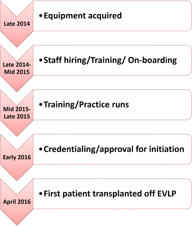
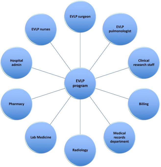
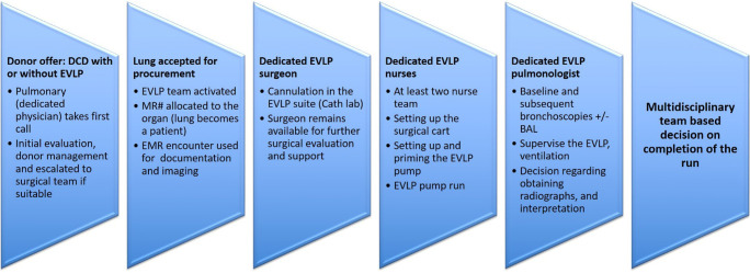
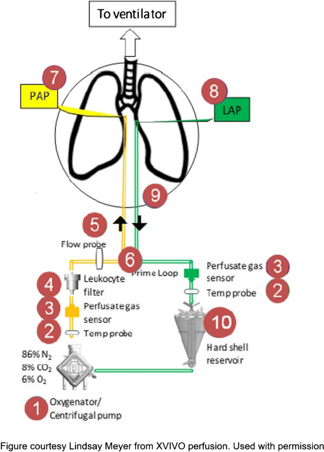
Lung transplantation is an established treatment for patients with end-stage lung disease. However, a shortage of donors, low lung utilization among potential donors, and waitlist mortality continue to be challenges. In the last decade, ex vivo lung perfusion (EVLP) has expanded the donor pool by allowing prolonged evaluation of marginal donor lungs and allowing reparative therapies for lungs, which are otherwise considered not transplantable. In this review, we describe in detail our experience with EVLP including our workflow, setup, operative technique, and protocols. Our multidisciplinary EVLP program functions with the collaboration of surgeons, pulmonologists, and EVLP nurses who run the pump. EVLP program has been a valuable addition to our program. Since Food and Drug Administration (FDA) approval in 2019, we experienced incremental increased lung transplant volume of 12% annually.

Keywords: Donor selection; Ex vivo lung perfusion; Lung transplantation.


Conflict of interest statement

Conflict of interestThe authors declare no competing interests.

Figures

Fig. 1
Fig. 1
The time line for EVLP program at our institution (EVLP, ex vivo lung perfusion)
Fig. 2
Fig. 2
The multidisciplinary EVLP team (EVLP, ex vivo lung perfusion)
Fig. 3
Fig. 3
Typical workflow at our institution (MR#, medical record number; EMR, electronic medical record; BAL, bronchoalveolar lavage; DCD, donation after circulatory death; EVLP, ex vivo lung perfusion)
Fig. 4
Fig. 4
The EVLP circuit (EVLP, ex vivo lung perfusion; PAP, pulmonary artery prime; LAP, left atrial prime)
Fig. 5
Fig. 5
A The organ chamber is set up. B The back table with all instruments for cannulation
Fig. 6
Fig. 6
A XPS LA cannula trimmed to size. B XPS LA cannula sutured to LA cuff. C Finished anastomosis (XPS, XVIVO® perfusion system; LA, left atrial)
Fig. 7
Fig. 7
A Types of XPS PA cannulas. B XPS cannula sutured to PA cuff. C Completed LA and PA anastomoses. D If the native PA is recovered in the donor lung, then the yellow cannula is used to cannulate PA and secured with silk ties (XPS, XVIVO® perfusion system; PA, pulmonary artery; LA, left atrial)
Fig. 8
Fig. 8
A Trachea is clamped with a Satinsky clamp and the stapled line removed. B Endotracheal tube inserted. C The ET tube is secured with silk ties and umbilical tape and ET tube clamped (remove the Satinsky clamp). D Retrograde flush with 1 l of Perfadex solution (ET, endotracheal)
Fig. 9
Fig. 9
A The circuit divided between clamps. B LA line (green) proximal to the bridge is clamped and second clamp on distal PA tubing (yellow). Retrograde flush from LA to PA done. C LA cannula connected. D PA tubing de-aired and PA cannula connected. E PA cannula removed. F LA clamp removed and placed on the bridge thus initiating the flow (LA, left atrial; PA, pulmonary artery)
Fig. 10
Fig. 10
A ET tube is clamped during rewarming. B Ventilation started when the LA temperature reaches 32 °F. C Bronchoscopy being performed
Fig. 11
Fig. 11
CXR of a successful EVLP run (CXR, chest X-ray; EVLP, ex vivo lung perfusion)
Fig. 12
Fig. 12
A Steen leak seen on bronchoscopy. B Extensive airway damage. C A tear in the bronchus in a DCD patient, which was missed on initial bronchoscopy but detected on EVLP assessments. D Progressive bogginess in the right lung with Steen and purulent secretions through ET tube. E CXR of the same lungs showing extensive consolidation in the right lung (EVLP, ex vivo lung perfusion; DCD, donation after circulatory death; ET, endotracheal)
Fig. 13
Fig. 13
Sequence of decannulation. A Both LA and PA lines clamped, ET tube clamped. B Cannulas disconnected. C Retrograde perfusion started (LA, left atrial; PA, pulmonary artery; ET, endotracheal)
Fig. 14
Fig. 14
A PA cannula disconnected. B LA cannula disconnected. C Satinsky clamp of trachea and ET tube removed. D Trachea stapled with TX-30 green stapler (Ethicon PROXIMATE TX Reloadable Linear Stapler) (PA, pulmonary artery; LA, left atrial; ET, endotracheal)
Fig. 15
Fig. 15
A Single lung EVLP run. B CXR after 3 h showing dense consolidation in the lower lobe (EVLP, ex vivo lung perfusion; CXR, chest X-ray)

References

    1. Organ Procurement and Transplantation Network (OPTN). Data as of Dec 31, 2020. Available at https://optn.transplant.hrsa.gov. Accessed 1 Feb 2021
    1. Charles EJ, Huerter ME, Wagner CE, Sharma AK, Zhao Y, Stoler MH, Mehaffey JH, Isbell JM, Lau CL, Tribble CG, Laubach VE, Kron IL. Donation after circulatory death lungs transplantable up to six hours after ex vivo lung perfusion. Ann Thorac Surg. 2016;102:1845–1853. doi: 10.1016/j.athoracsur.2016.06.043. - DOI - PMC - PubMed
    1. Reeb J, Keshavjee S, Cypel M. Expanding the lung donor pool: advancements and emerging pathways. Curr Opin Organ Transplant. 2015;20:498–505. doi: 10.1097/MOT.0000000000000233. - DOI - PubMed
    1. Valapour M, Lehr CJ, Skeans MA, Smith JM, Uccellini K, Goff R, Foutz J, Israni AK, Snyder JJ, Kasiske BL. OPTN/SRTR 2018 annual data report: lung. Am J Transplant. 2020;20:427–508. doi: 10.1111/ajt.15677. - DOI - PubMed
    1. Chaney J, Suzuki Y, Cantu E, 3rd, van Berkel V. Lung donor selection criteria. J Thorac Dis. 2014;6:1032–1038. - PMC - PubMed

LinkOut - more resources