. 2020 Dec 1;30(49):2005029.
doi: 10.1002/adfm.202005029. Epub 2020 Sep 9.

Light: A Magical Tool for Controlled Drug Delivery

Affiliations

Light: A Magical Tool for Controlled Drug Delivery

Yu Tao et al. Adv Funct Mater. .

Abstract

Light is a particularly appealing tool for on-demand drug delivery due to its noninvasive nature, ease of application and exquisite temporal and spatial control. Great progress has been achieved in the development of novel light-driven drug delivery strategies with both breadth and depth. Light-controlled drug delivery platforms can be generally categorized into three groups: photochemical, photothermal, and photoisomerization-mediated therapies. Various advanced materials, such as metal nanoparticles, metal sulfides and oxides, metal-organic frameworks, carbon nanomaterials, upconversion nanoparticles, semiconductor nanoparticles, stimuli-responsive micelles, polymer- and liposome-based nanoparticles have been applied for light-stimulated drug delivery. In view of the increasing interest in on-demand targeted drug delivery, we review the development of light-responsive systems with a focus on recent advances, key limitations, and future directions.

Keywords: Light; drug delivery; photochemical; photoisomerization; photothermal.


Figures

Figure 1.
Figure 1.
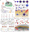
Schematic illustration of light-controlled drug delivery.
Figure 2.
Figure 2.
(A) Schematic illustration of ROS cascade-responsive drug release of PEG-stearamine conjugate nanomicelles loaded with DOX and pheophorbide A for enhanced regional chemo-photodynamic therapy. Reproduced with permission.[49] Copyright 2020, Elsevier. (B) Synthesis of NIR photoactivatable semiconducting polymer nanoblockaders for metastasis-inhibited cancer therapy. Reproduced with permission.[50] Copyright 2019, Wiley-VCH. (C) Chemical structure and self-assembly of NIR photolabile semiconducting polymer nanotransducer. Schematic of photolabile semiconducting polymer nanotransducer-mediated delivery and photoregulation of CRISPR/Cas9 gene editing under NIR light irradiation. Reproduced with permission.[53] Copyright 2019, Wiley-VCH.
Figure 3.
Figure 3.
(A) Schematic illustration of the structure of tirapazamine/upconversion nanoparticle@porphyrinic MOFs and their application to tumor treatment through a combination of NIR light-triggered photodynamic therapy and hypoxia-activated chemotherapy with immunotherapy. Reproduced with permission.[66] Copyright 2020, American Chemical Society. (B) Schematic illustrations of synthesis of upconversion nanocapsules, and folate receptor-mediated cellular uptake and NIR modulated intracellular siRNA delivery and therapy. Reproduced with permission.[67] Copyright 2018, Elsevier. (C) Schematic illustration of an upconversion-nanoparticle-centered Au nanoparticles tetrahedron used for senescence clearence. Reproduced with permission.[72] Copyright 2020, Wiley-VCH.
Figure 4.
Figure 4.
(A) Schematic representation of selenium-inserted polymeric nanoparticles with immediate drug release and highly efficient cytoplasmic translocation features upon NIR irradiation for combined chemo-photothermotherapy. Reproduced with permission.[78] Copyright 2017, American Chemical Society. (B) Formation and mechanism of the light-activated shrinkable nanoplatform. Reproduced with permission.[79] Copyright 2018, American Chemical Society. (C) Schematic of how the photo-triggered nanoplatforms worked in combination with hypoxia-triggered and photodynamic therapy strategy. Reproduced with permission.[81] Copyright 2018, American Chemical Society.
Figure 5.
Figure 5.
A brief history of the application of nanomaterials for photothermally controlled drug release.
Figure 6.
Figure 6.
(A) Schematic illustration of synergistic anticancer system combining photothermal treatment and inflammation-mediated active targeting chemotherapy. (B) The synergistic therapeutic efficiency of photothermal therapy and inflammation-mediated active targeting chemotherapy. The growth profiles of tumors in mice that received different treatments. The HE staining and immunohistochemical staining (caspase-3) of tumor sections after administration of different treatments. Reproduced with permission.[104] Copyright 2019, Wiley-VCH. (C) Rational design and applications of Se@carbon nanotubes in cancer chemo-photothermal therapy. (D) T2-weighted magnetic resonance images of MDA-MB-231 tumor-bearing mice after various treatments for 14 days. Reproduced with permission.[116] Copyright 2018, Wiley-VCH.
Figure 7.
Figure 7.
(A) Schematic of membrane fusion and coating. Membrane materials are derived from red blood cells and B16-F10 cells and then fused together. The resulting hybrid membrane is used to camouflage DOX-loaded hollow copper sulfide nanoparticles to produce nanoparticles for synergistic photothermal/chemotherapy of melanoma. Reproduced with permission.[120] Copyright 2018, American Chemical Society. (B) Synthetic process and therapeutic mechanism of BSA-IrO2 NPs. Temperature-dependent H2O2-triggered O2 generation/consumption of 1,3-diphenylisobenzofuran under irradiation/concentration-dependent photothermal curves by BSA-IrO2 NPs. In vitro photoacoustic images and photoacoustic signals of BSA-IrO2 NPs at different concentrations of Ir and upon the addition of H2O2, as well as CT images and Hounsfield unit values of BSA-IrO2 NPs solutions. Reproduced with permission.[124] Copyright 2018, Wiley-VCH. (C) Illustration of atomic-level nanorings, a theranostic agent for photoacoustic imaging, photodynamic therapy and photothermal therapy for cancer. Reproduced with permission.[128] Copyright 2020, American Chemical Society. (D) Schematic illustration for the synthesis and underlying mechanisms of tungsten-based polyoxometalate nanoclusters for non-inflammatory photothermal therapy with rapid renal clearance. Reproduced with permission.[144] Copyright 2020, American Chemical Society.
Figure 8.
Figure 8.
(A) Schematic illustration of the preparation of L1057 NPs as a theranostic agent. Reproduced with permission.[158] Copyright 2020, American Chemical Society. (B) Schematic illustration of polymer-dispersed liquid crystal nanoparticles for chemo-photothermal treatment for Aβ aggregation. Reproduced with permission.[160] Copyright 2020, Wiley-VCH. (C) Schematic illustration of Cy7–2-dicyanomethylene-3-cyano-4,5,5-trimethyl-2,5-dihydrofuran self-assembly into a nanodisc to exhibit the phototheranostic functions and accumulation at tumor site, thereby improving the imaging-guided cancer therapeutic efficacy in vivo. Reproduced with permission.[174] Copyright 2020, Wiley-VCH. (D) Schematic illustration of fabrication of eukaryotic-prokaryotic vesicles coated poly(lactic-co-glycolic acid)-indocyanine green moiety-implanted eukaryotic-prokaryotic vesicle nanovaccine. Survival curves of mice after various treatments. Flow cytometric analysis of PE-labeled T-Select MHC Tetramer-positive cells gated CD8+ cells expressed in the therapeutic efficacy trial. Reproduced with permission.[175] Copyright 2020, Wiley-VCH.
Figure 9.
Figure 9.
(A) Synthesis of light-responsive azobenzene-functionalized covalent organic framework (TTA-AzoDFP) obtained under solvothermal conditions. Schematic representation of the release of Rhodamine B from the pores of TTA-AzoDFP after UV irradiation, UV-Vis generated release profile of Rhodamine B-loaded TTA-AzoDFP upon UV irradiation. Reproduced with permission.[205] Copyright 2019, American Chemical Society. (B) Chemical structures of phototrexate in the trans and cis configurations. Zoomed views of cis (in purple) phototrexate superimposed on the crystallographic structure of methotrexate (in orange) in the active site of the human dihydrofolate reductase. HeLa cell viability assay at different concentrations of methotrexate, cis-phototrexate and trans-phototrexate. Reproduced with permission.[223] Copyright 2018, American Chemical Society. (C) Schematic presentations of sequential self-assembly preparation of photosensitizer-containing copolymer ternary polyplexes, and light-induced reversible 1O2 generation. Reproduced with permission.[226] Copyright 2019, American Chemical Society. (D) Schematic illustration of synthetic light-driven substrate channeling for precise regulation of enzyme cascade activity on DNA origami with different wavelength of light. Reproduced with permission.[228] Copyright 2018, American Chemical Society.
Figure 10.
Figure 10.
(A) Schematic illustration of UCNPs-DNAAzo/DOX assembly and UCNPs-DNAAzo/DOX-TAT-hyaluronic acid synthesis. Reproduced with permission.[240] Copyright 2019, Wiley-VCH. (B) Illustration of the fabrication of the up/downconversion nanoparticle functionalized hollow polymer nanocapsules and the NIR light induced decomposing process from 180 nm nanocapsules to scattered polymers and 20 nm up/downconversion nanoparticles. Reproduced with permission.[241] Copyright 2018, Wiley-VCH. (C) Schematic illustration of NIR-driven reversible bacteria clustering. Reproduced with permission.[243] Copyright 2019, Elsevier. (D) Schematic illustration of NIR-controlled cage mimicking system for Curcumin delivery through spiropyran modified on mesoporous silica coated upconvertion nanoparticles. The Curcumin release by taking advantage of the upconversion capability of UCNP and the conformational transformation of spiropyran molecules immobilized on mesoporous silica. Reproduced with permission.[245] Copyright 2017, Elsevier.
Figure 11.
Figure 11.
Applications of light-controlled drug delivery.

References

    1. Paull R, Wolfe J, Hébert P, Sinkula M, Nat. Biotechnol 2003, 21, 1144. - PubMed
    1. Roco MC, Curr. Opin. Biotechnol 2003, 14, 337; - PubMed
    2. Sahoo SK, Parveen S, Panda JJ, Nanomedicine: NBM 2007, 3, 20. - PubMed
    1. Karimi M, Bahrami S, Ravari SB, Zangabad PS, Mirshekari H, Bozorgomid M, Shahreza S, Sori M, Hamblin MR, Expert Opin. Drug Deliv 2016, 13, 1609; - PMC - PubMed
    2. Wang H, Ke F, Mararenko A, Wei Z, Banerjee P, Zhou S, Nanoscale 2014, 6, 7443; - PubMed
    3. Robinson JT, Tabakman SM, Liang Y, Wang H, Sanchez Casalongue H, Vinh D, Dai H, J. Am. Chem. Soc 2011, 133, 6825; - PubMed
    4. Karimi M, Solati N, Amiri M, Mirshekari H, Mohamed E, Taheri M, Hashemkhani M, Saeidi A, Estiar MA, Kiani P, Ghasemi A, Basri SMM, Aref AR, Hamblin MR, Expert Opin. Drug Deliv 2015, 12, 1071; - PMC - PubMed
    5. Liu JL, Dixit AB, Robertson KL, Qiao E, Black LW, Proc. Natl. Acad. Sci. USA 2014, 111, 13319; - PMC - PubMed
    6. Hosseinidoust Z, Mostaghaci B, Yasa O, Park B-W, Singh AV, Sitti M, Adv. Drug Delivery Rev 2016, 106, 27; - PubMed
    7. Karimi M, Eslami M, Sahandi-Zangabad P, Mirab F, Farajisafiloo N, Shafaei Z, Ghosh D, Bozorgomid M, Dashkhaneh F, Hamblin MR, Wiley Interdiscip. Rev.: Nanomed. Nanobiotechnol 2016, 8, 696; - PMC - PubMed
    8. Karimi M, Sahandi Zangabad P, Ghasemi A, Amiri M, Bahrami M, Malekzad H, Ghahramanzadeh Asl H, Mahdieh Z, Bozorgomid M, Ghasemi A, Rahmani Taji Boyuk MR, Hamblin MR, ACS Appl. Mater. Inter 2016, 8, 21107; - PMC - PubMed
    9. Wang L, Yuan Y, Lin S, Huang J, Dai J, Jiang Q, Cheng D, Shuai X, Biomaterials 2016, 78, 40; - PubMed
    10. Huang S, Liu J, He Q, Chen H, Cui J, Xu S, Zhao Y, Chen C, Wang L, Nano Research 2015, 8, 4038.
    1. Heo DN, Ko WK, Moon HJ, Kim HJ, Lee SJ, Lee JB, Bae MS, Yi JK, Hwang YS, Bang JB, Kim EC, Do SH, Kwon IK, ACS Nano 2014, 8, 12049; - PubMed
    2. Bensellam M, Laybutt DR, Jonas J-C, Mol. Cell. Endocrinol 2012, 364, 1; - PubMed
    3. Karimi M, Zare H, Bakhshian Nik A, Yazdani N, Hamrang M, Mohamed E, Sahandi Zangabad P, Moosavi Basri SM, Bakhtiari L, Hamblin MR, Nanomedicine 2016, 11, 513; - PMC - PubMed
    4. Carrasco E, Calvo MI, Blázquez-Castro A, Vecchio D, Zamarrón A, de Almeida IJD, Stockert JC, Hamblin MR, Juarranz Á, Espada J, J. Invest. Dermatol 2015, 135, 2611; - PMC - PubMed
    5. Abrahamse H, Hamblin MR, Biochem. J 2016, 473, 347; - PMC - PubMed
    6. Yu C, Avci P, Canteenwala T, Chiang LY, Chen BJ, Hamblin MR, J. Nanosci.Nanotechnol 2016, 16, 171. - PMC - PubMed
    1. Wang Y, Li B, Zhang L, Song H, Zhang L, ACS Appl. Mater. Inter 2012, 5, 11.

LinkOut - more resources