

The Age of Brain Organoids: Tailoring Cell Identity and Functionality for Normal Brain Development and Disease Modeling
- PMID: 34483818
- PMCID: PMC8414411
- DOI: 10.3389/fnins.2021.674563
The Age of Brain Organoids: Tailoring Cell Identity and Functionality for Normal Brain Development and Disease Modeling
Abstract
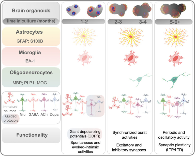
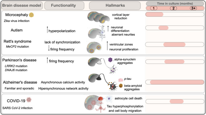
Over the past years, brain development has been investigated in rodent models, which were particularly relevant to establish the role of specific genes in this process. However, the cytoarchitectonic features, which determine neuronal network formation complexity, are unique to humans. This implies that the developmental program of the human brain and neurological disorders can only partly be reproduced in rodents. Advancement in the study of the human brain surged with cultures of human brain tissue in the lab, generated from induced pluripotent cells reprogrammed from human somatic tissue. These cultures, termed brain organoids, offer an invaluable model for the study of the human brain. Brain organoids reproduce the cytoarchitecture of the cortex and can develop multiple brain regions and cell types. Integration of functional activity of neural cells within brain organoids with genetic, cellular, and morphological data in a comprehensive model for human development and disease is key to advance in the field. Because the functional activity of neural cells within brain organoids relies on cell repertoire and time in culture, here, we review data supporting the gradual formation of complex neural networks in light of cell maturity within brain organoids. In this context, we discuss how the technology behind brain organoids brought advances in understanding neurodevelopmental, pathogen-induced, and neurodegenerative diseases.
Keywords: SARS-CoV-2; Zika virus; brain development; brain organoids; electrophysiology; human pluripotent stem cells (hPSC); neurodegenerative diseases; neurodevelopmental disorders.
Copyright © 2021 Porciúncula, Goto-Silva, Ledur and Rehen.
Conflict of interest statement
The authors declare that the research was conducted in the absence of any commercial or financial relationships that could be construed as a potential conflict of interest.
Figures
References
-
- Alić I., Goh P. A., Murray A., Portelius E., Gkanatsiou E., Gough G., et al. (2020). Patient-specific Alzheimer-like pathology in trisomy 21 cerebral organoids reveals BACE2 as a gene dose-sensitive AD suppressor in human brain. Mol. Psychiatry 10, 1–23. 10.1038/s41380-020-0806-5 - DOI - PMC - PubMed
Publication types
LinkOut - more resources
Full Text Sources
Miscellaneous