

Incidental discovery of duplicated inferior vena cava in a septuagenarian: the radiologist's viewpoint
- PMID: 34484518
- PMCID: PMC8405935
- DOI: 10.1016/j.radcr.2021.07.070
Incidental discovery of duplicated inferior vena cava in a septuagenarian: the radiologist's viewpoint
Erratum in
-
Erratum regarding missing patient consent statements in previously published articles.Radiol Case Rep. 2022 Nov 25;18(2):730-731. doi: 10.1016/j.radcr.2022.10.049. eCollection 2023 Feb. Radiol Case Rep. 2022. PMID: 36588598 Free PMC article.
Abstract
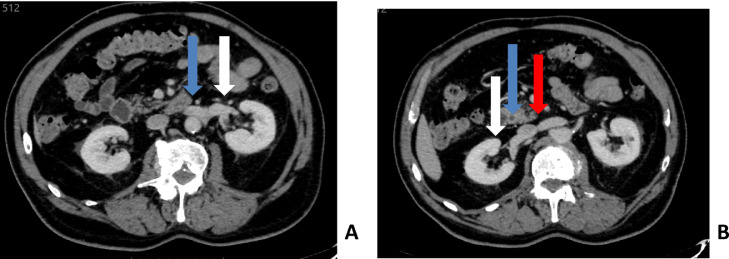
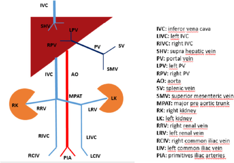
Duplication of the inferior vena cava is a rare malformation, normally without clinical impact, explained by abnormal development and regression of certain segments of the venous system during embryonic life. However, its presence and type should be systematically reported in the radiological report because of its potential implications for diagnostic and interventional procedures. This observation describes the case of a 77-year-old man with a complete asymmetric duplication of the inferior vena cava (type III IVC according to Natsis) that was incidentally discovered on CT-scan.
Keywords: Duplication; Imaging; Inferior vena cava.
© 2021 The Authors. Published by Elsevier Inc. on behalf of University of Washington.
Figures


References
-
- Inferior vena cava. Available at: https://en.wikipedia.org/wiki/Inferior_vena_cava. Accessed on July 2021.
-
- Natsis K, Apostolidis S, Noussios G. Duplication of the inferior vena cava: anatomy, embryology and classification proposal. Anat Sci Int. 2010;85(1):56–60. - PubMed
-
- Yagel S, Kivilevitch Z, Cohen SM. The fetal venous system, part I: normal embryology, anatomy, hemodynamics, ultrasound evaluation and Doppler investigation. Ultrasound Obstet Gynecol. 2010;35:741–750. - PubMed
Publication types
LinkOut - more resources
Full Text Sources
