Case Reports
. 2021 Aug 26;16(11):3300-3303.
doi: 10.1016/j.radcr.2021.07.080. eCollection 2021 Nov.

Intraosseous metaplastic meningioma: A case report

Affiliations
Case Reports

Intraosseous metaplastic meningioma: A case report

Yusuke Utsunomiya et al. Radiol Case Rep. .

Erratum in

Abstract

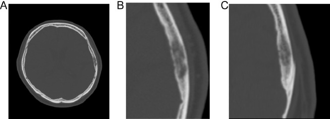
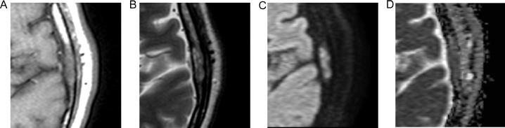
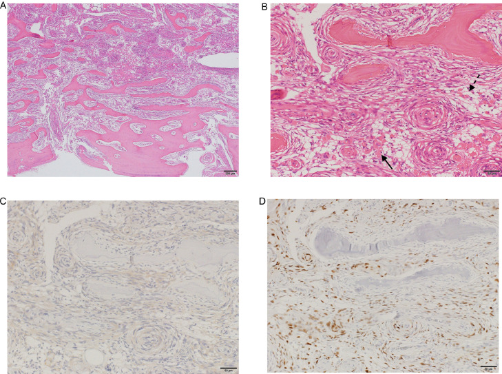
Metaplastic meningioma is a rare World Health Organization Grade I meningioma subtype, accounting for 0.2%-1.6% of all meningiomas. Primary extradural meningiomas represent less than 2% of all meningiomas, with intraosseous meningioma as a subtype of primary extradural meningiomas. Herein, we report the case of a 65-year-old male presenting with headache. His computed tomography scans showed an osteolytic left parietal bone mass, and magnetic resonance imaging revealed hyperintense dots in the mass on T1-weighted images. The mass was then resected and diagnosed on histopathological examination as an intraosseous metaplastic meningioma.

Keywords: Calvarial; Imaging; Intraosseous; Meningioma; Metaplastic.


Figures

Fig. 1 –
Fig. 1
Head computed tomography of a 65-year-old male presenting with headache. Axial (A, B) and coronal (C) images show a 19-mm-long, well-defined, left parietal bone mass near the squamosal suture. The main part of the mass is located in a diploic space, and the trabeculae are sparse but well preserved. There is also a mild bony bulge, although there are no tears in the inner or outer plates.
Fig. 2 –
Fig. 2
Magnetic resonance images of the mass. Axial T1-weighted image (A) shows the mass is isointense relative to muscles and includes internal hyperintense dots. Axial T2-weighted image (B) shows heterogeneous intensity. Axial diffusion-weighted image (C) and the corresponding apparent diffusion coefficient map (D) show diffusion restriction.
Fig. 3 –
Fig. 3
Histopathological examination of the mass reveals tumor growth in the intertrabecular space and whorl patterns of spindle-shaped cells (hematoxylin-eosin stain, original magnification, ×4 [A] and ×20 [B]). The tumor is marked with ossification (arrow) and lipomatous tissue (dashed arrow) inclusions, which are different from the pre-existing bone trabeculae and bone marrow fat. Immunohistochemistry shows that the tumor cells express epithelial membrane antigen (C) and progesterone receptor (D), but they are negative for MIB-1 (not shown here). Given these findings, the final diagnosis is intraosseous metaplastic meningioma (World Health Organization Grade I).

References

    1. Kim T., Kim J.W., Ji S.Y., Kang H., Kim K.-M., Kim Y.H. Intracranial metaplastic meningioma: clinical and radiological characteristics of 11 cases. J Korean Neurosurg Soc. 2020;63:657–663. doi: 10.3340/jkns.2020.0151. - DOI - PMC - PubMed
    1. Wang D.J., Xie Q., Gong Y., Mao Y., Wang Y., Cheng H.X. Histopathological classification and location of consecutively operated meningiomas at a single institution in China from 2001 to 2010. Chin Med J (Engl). 2013;126:488–493. doi: 10.3760/cma.j.issn.0366-6999.20122874. - DOI - PubMed
    1. Zouaoui S., Darlix A., Rigau V., Mathieu-Daudé H., Bauchet F., Bessaoud F. Descriptive epidemiology of 13,038 newly diagnosed and histologically confirmed meningiomas in France: 2006–2010. Neurochirurgie. 2018;64:15–21. doi: 10.1016/j.neuchi.2014.11.013. - DOI - PubMed
    1. Bhat A.R., Wani M.A., Kirmani A.R., Ramzan A.U. Histological-subtypes and anatomical location correlated in meningeal brain tumors (meningiomas) J Neurosci Rural Pract. 2014;5:244–249. doi: 10.4103/0976-3147.133568. - DOI - PMC - PubMed
    1. Tang H., Sun H., Chen H., Gong Y., Mao Y., Xie Q. Clinicopathological analysis of metaplastic meningioma: report of 15 cases in Huashan Hospital. Chinese J Cancer Res. 2013;25:112–118. doi: 10.3978/j.issn.1000-9604.2013.01.10. - DOI - PMC - PubMed

Publication types

LinkOut - more resources