Case Reports
. 2021 Aug 26;16(11):3315-3320.
doi: 10.1016/j.radcr.2021.07.072. eCollection 2021 Nov.

Intra-articular hip joint osteoid osteoma: Challenging diagnosis and percutaneous radiofrequency ablation treatment

Affiliations
Case Reports

Intra-articular hip joint osteoid osteoma: Challenging diagnosis and percutaneous radiofrequency ablation treatment

Talal Al Kuhaimi et al. Radiol Case Rep. .

Erratum in

Abstract

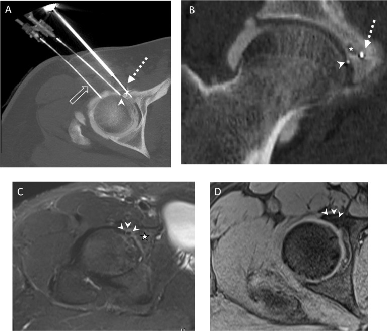
Atypical intra-articular osteoid osteoma can be difficult to diagnose and challenging to treat. We report a case of a right acetabular subchondral intra-articular osteoid osteoma in a young male patient which was initially diagnosed as femoroacetabular impingement due to its atypical clinical and radiological presentations. After fully working up the patient the lesion was successfully treated with percutaneous CT-guided low-power bipolar radiofrequency ablation using several per procedural articular cartilage thermal protective measures including intra-articular thermocouple, and continuous per procedural joint space cooling with Dextrose 5% solution. A precise RFA electrode placement, using the No-touch technique, and applying different passive and active thermal protective measures were helpful in avoiding collateral damage of the hip joint articular cartilages. atypical intra-articular osteoid osteomas necessitate pertinent correlation between the clinical and radiological presentations. As far as intra-articular or subchondral nidus ablation is concerned, thermal protective measures should be considered.

Keywords: Bipolar Radiofrequency ablation; Hip joint; Intra-articular; Osteoid osteoma.


Figures

Fig 1
Fig. 1
(a) Plain radiograph of right hip demonstrates normal hip joint space without osteochondral lesion, or reactive sclerosis. (b) CT scan of right hip demonstrates subchondral lucent lesion (solid arrow) of the anterior acetabular wall without reactive sclerosis or periosteal reaction. (c d) Coronal and axial T2 fat-saturated weighted MR images demonstrate high signal intensity nidus (white open arrow) with surrounding reactive bone marrow edema (white solid arrow). No joint effusion is noted. No injectate fills the subchondral defect. (e) Axial T1 fat-saturated MR Arthrogram image demonstrates normal overlying cartilage without delamination. The subchondral defect is not filled with injectate (arrow heads). (f), Axial subtraction post contrast MR image demonstrates avidly enhancing subchondral nidus (arrow head) with mild perilesional bone marrow enhancement (white solid arrow). (Color version of the figure is available online.)
Fig 2
Fig. 2
(a b) Axial and coronal oblique intra procedural CT scan images, demonstrating placement of low energy bipolar RFA electrode (dotted white arrow) anterior and medial to the osteoid osteoma nidus (white star) using No-touch technique. The thermocouple (white arrowhead) is intra-articularly placed and seated posterior and lateral to the nidus. Intra-articular hydrodissection and cooling with cold dextrose mixed with omnipaque were performed through the spinal needle (open white arrow). (c) Post treatment axial oblique T2 fat-saturated MR image demonstrates low signal intensity nidus with improvement of the surrounding edema and no cartilage damage. (d) Post treatment axial T1 fat-saturated dynamic enhanced MR image demonstrates no enhancing nidus.

References

    1. Burrows HJ. Osteoid Osteoma. Proceedings of the Royal Society of Medicine. 1953;46(12):29-34. doi:10.1177/003591575304601203. - DOI - PMC - PubMed
    1. Nehme AH, Bou Ghannam AG, Imad JP, Jabbour FC, Moucharafieh R, Wehbe J. Arthroscopic Excision of Intra-Articular Hip Osteoid Osteoma: A Report of 2 Cases. Case Reports in Orthopedics. 2012. 2012:1–4. doi: 10.1155/2012/820501. - DOI - PMC - PubMed
    1. Edeiken J, DePalma AF, Hodes PJ. Osteoid osteoma. (Roentgenographic emphasis) Clin Orthop Relat Res. 1966;49:201–206. - PubMed
    1. Carter TR. Osteoid osteoma of the hip: an alternate method of excision. Orthop Rev. 1990;19(10):903–905. - PubMed
    1. Bettelli G, Capanna R, Van Horn J, Ruggieri P, Biagini R, Campanacci M. Osteoid Osteoma and Osteoblastoma of the Pelvis. Clinical Orthopaedics and Related Research. 1989;(247) doi: 10.1097/00003086-198910000-00036. &NA. - DOI - PubMed

Publication types