Review
. 2021 Aug 30:2021:4936571.
doi: 10.1155/2021/4936571. eCollection 2021.

COVID-19 and Acute Coronary Syndromes: From Pathophysiology to Clinical Perspectives

Affiliations
Review

COVID-19 and Acute Coronary Syndromes: From Pathophysiology to Clinical Perspectives

Luca Esposito et al. Oxid Med Cell Longev. .

Abstract

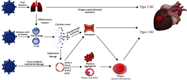
Acute coronary syndromes (ACS) are frequently reported in patients with coronavirus disease 2019 (COVID-19) and may impact patient clinical course and mortality. Although the underlying pathogenesis remains unclear, several potential mechanisms have been hypothesized, including oxygen supply/demand imbalance, direct viral cellular damage, systemic inflammatory response with cytokine-mediated injury, microvascular thrombosis, and endothelial dysfunction. The severe hypoxic state, combined with other conditions frequently reported in COVID-19, namely sepsis, tachyarrhythmias, anemia, hypotension, and shock, can induce a myocardial damage due to the mismatch between oxygen supply and demand and results in type 2 myocardial infarction (MI). In addition, COVID-19 promotes atherosclerotic plaque instability and thrombus formation and may precipitate type 1 MI. Patients with severe disease often show decrease in platelets count, higher levels of d-dimer, ultralarge von Willebrand factor multimers, tissue factor, and prolongation of prothrombin time, which reflects a prothrombotic state. An endothelial dysfunction has been described as a consequence of the direct viral effects and of the hyperinflammatory environment. The expression of tissue factor, von Willebrand factor, thromboxane, and plasminogen activator inhibitor-1 promotes the prothrombotic status. In addition, endothelial cells generate superoxide anions, with enhanced local oxidative stress, and endothelin-1, which affects the vasodilator/vasoconstrictor balance and platelet aggregation. The optimal management of COVID-19 patients is a challenge both for logistic and clinical reasons. A deeper understanding of ACS pathophysiology may yield novel research insights and therapeutic perspectives in higher cardiovascular risk subjects with COVID-19.


Conflict of interest statement

The authors declare that they have no conflicts of interest. The authors have no commercial or financial relationships that could be construed as a potential conflict of interest.

Figures

Figure 1
Figure 1
Pathophysiology of ACS in COVID-19. Mechanisms involved in the pathophysiology of ACS in patients with COVID-19. SARS-CoV-2, by binding the ACE2 receptors expressed on the surface of the host cell, may infect pneumocytes, macrophages, and endothelial cells. The respiratory impairment related to the pulmonary involvement, ranging from pneumonia to ARDS in severe forms, causes hypoxia and type 2 MI due to the oxygen supply/demand mismatch. Also, the infection promotes an aberrant inflammatory response resulting in the release of cytokines and proinflammatory molecules such as IL-1, IL-6, IL-7, TNFα, and IFNγ. Cytokines have the potential to damage the endothelial function with increased production of oxidative stress agents and prothrombotic factors. SARS-CoV-2 may also exert a direct cellular effect by interacting with molecules expressed on the surface of the endothelial cells. In turn, the inflammatory environment enhances the instability of preexisting atheromatous plaques, promotes platelets activation and aggregation, and upregulates the sympathetic nervous system resulting in increased vasomotility and coronary spasm. The interplay of all these mechanisms may favor plaque rupture and thrombosis leading to type 1 MI. ACS: acute coronary syndrome; ACE2: angiotensin-converting enzyme 2; COVID-19: coronavirus disease 2019; IFNγ: interferon γ; IL-1: interleukin 1; IL-6: interleukin 6; IL-7: interleukin 7; MI: myocardial infarction; SARS-CoV-2: severe acute respiratory syndrome coronavirus 2; TNFα: tumor necrosis factor α.

References

    1. Guan W. J., Ni Z. Y., Hu Y., et al. Clinical characteristics of coronavirus disease 2019 in China. The New England journal of medicine. 2020;382(18):1708–1720. doi: 10.1056/NEJMoa2002032. - DOI - PMC - PubMed
    1. Progress report on the coronavirus pandemic. Nature. 2020;584 doi: 10.1038/d41586-020-02414-1. - DOI - PubMed
    1. Silverio A., di Maio M., Citro R., et al. Cardiovascular risk factors and mortality in hospitalized patients with COVID-19: systematic review and meta-analysis of 45 studies and 18,300 patients. BMC cardiovascular disorders. 2021;21(1):p. 23. doi: 10.1186/s12872-020-01816-3. - DOI - PMC - PubMed
    1. Shi S., Qin M., Shen B., et al. Association of cardiac injury with mortality in hospitalized patients with COVID-19 in Wuhan, China. JAMA cardiology. 2020;5(7):802–810. doi: 10.1001/jamacardio.2020.0950. - DOI - PMC - PubMed
    1. Citro R., Pontone G., Bellino M., et al. Role of multimodality imaging in evaluation of cardiovascular involvement in COVID-19. Trends in cardiovascular medicine. 2021;31(1):8–16. doi: 10.1016/j.tcm.2020.10.001. - DOI - PMC - PubMed