

A retrospective cone beam computed tomography analysis of cemento-osseous dysplasia
- PMID: 34484583
- PMCID: PMC8403794
- DOI: 10.1016/j.jds.2021.03.009
A retrospective cone beam computed tomography analysis of cemento-osseous dysplasia
Abstract
Background/purpose: Radiological examination is indispensable in the diagnosis and follow-up of cemento-osseous dysplasia (COD). The aim of this retrospective study was to describe a series of COD cases, identify the frequencies of COD subtypes, and investigate the demographic and radiological characteristics in relation to subtypes.
Materials and methods: Cone beam computed tomography (CBCT) images/reports of patients with a diagnosis of COD were included in the study. The data collected included information on the age, sex, subtype of COD, location of COD, and region involved. Information regarding the internal density, effects on surrounding structures, and presence of concomitant lesions was also collected. The data obtained were evaluated statistically.
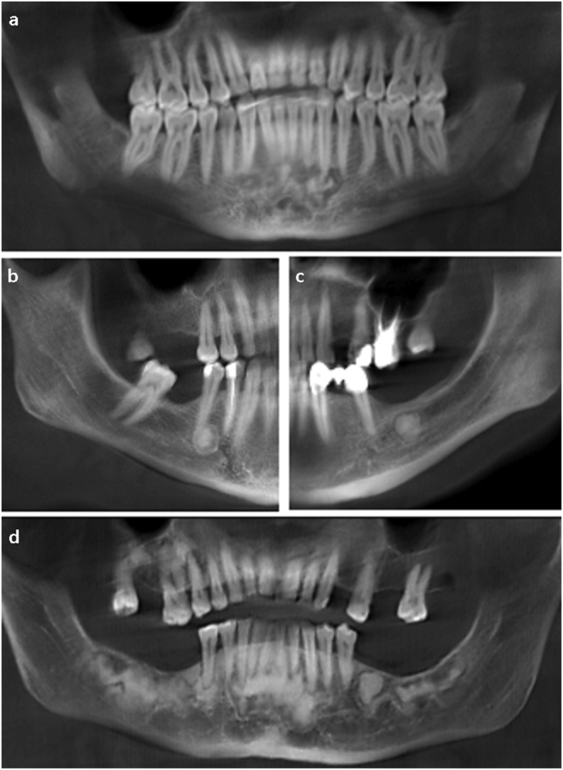
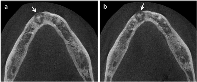
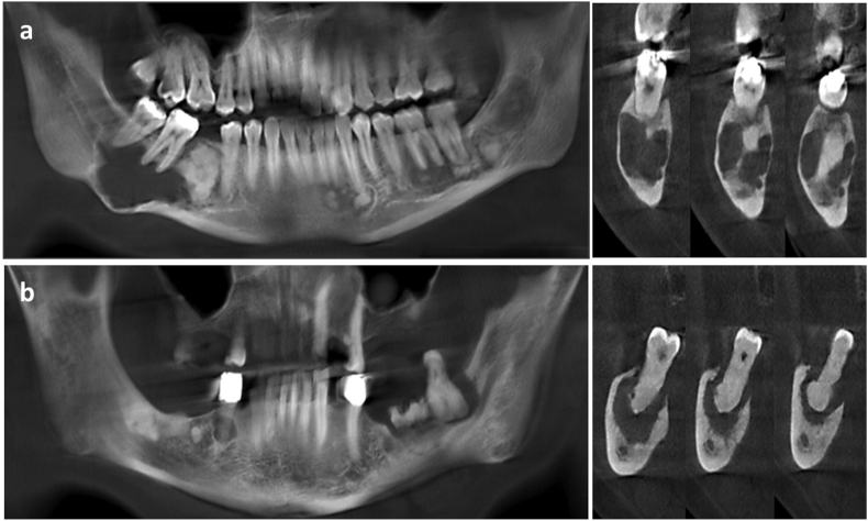
Results: The study group included CBCT images of 142 patients (130 females (91.5%) and 12 males (8.5%)) with a mean age of 46.97 ± 10.57 years. The mandible was involved in almost all cases (99.3%). The most common subtype was florid COD (51.4%) and lesions with hyperdense internal density (81.7%) were more commonly observed. Cortical thinning (78.2%) was a prominent feature. The frequency of root resorption in periapical COD cases (57.1%) was observed to be significantly higher (p < 0.05). All hypercementosis cases were associated with florid subtype (p < 0.05). In a minority of cases (6.3%), the lesions were associated with bone cysts and osteomyelitis.
Conclusion: CBCT images clearly demonstrated the effect of COD lesions on surrounding structures. CBCT is an appropriate imaging modality for the diagnosis and follow-up of COD which is the most common fibro-osseous lesion in clinical practice.
Keywords: Cemento-osseous dysplasia; Cone beam computed tomography; Florid; Subtype.
© 2021 Association for Dental Sciences of the Republic of China. Publishing services by Elsevier B.V.
Conflict of interest statement
The authors have no conflicts of interest relevant to this article.
Figures
References
-
- El-Naggar A.K., Chan J.K.C., Grandis J.R., Takata T., Slootweg P.J., editors. WHO classification of head and neck tumours. 4th ed. IARC Press; Lyon, France: 2017. pp. 251–255.
-
- Cavalcanti P.H.P., Nascimento E.H.L., Pontual M.L.D.A. Cemento-osseous dysplasias: imaging features based on cone beam computed tomography scans. Braz Dent J. 2018;29:99–104. - PubMed
-
- Oh D., Samuels J., Chaw S. Cemento-osseous dysplasia: Re-visited. J Dent Oral Health. 2019;1:1–11.
-
- Kato C.N.A.O., Barra S.G., Amaral T.M.P. Cone-beam computed tomography analysis of cemento-osseous dysplasia-induced changes in adjacent structures in a Brazilian population. Clin Oral Invest. 2020;24:2899–2908. - PubMed
-
- Waldron C.A. Fibro-osseous lesions of the jaws. J Oral Maxillofac Surg. 1993;51:828–835. - PubMed
LinkOut - more resources
Full Text Sources
