

Biomaterials Used for Suture Anchors in Orthopedic Surgery
- PMID: 34484619
- PMCID: PMC8380519
- DOI: 10.4055/cios20317
Biomaterials Used for Suture Anchors in Orthopedic Surgery
Abstract
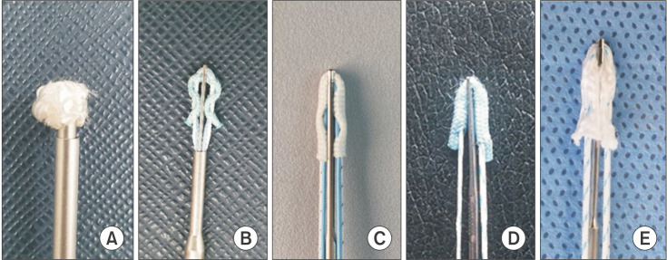
Suture anchors are broadly used for attaching soft tissue (e.g., tendons, ligaments, and meniscus) to the bone and have become essential devices in sports medicine and during arthroscopic surgery. As the usage of suture anchors has increased, various material-specific advantages and challenges have been reported. As a result, suture anchors are continually changing to become safer and more efficient. In this ever-changing environment, it is clinically essential for the surgeon to understand the key characteristics of existing anchors sufficiently. This paper aims to summarize the current concepts on the characteristics of available suture anchors.
Keywords: Arthroscopy; Biocompatible materials; Knee; Shoulder; Sports medicine; Suture anchor.
Copyright © 2021 by The Korean Orthopaedic Association.
Conflict of interest statement
CONFLICT OF INTEREST: No potential conflict of interest relevant to this article was reported.
Figures
Similar articles
-
Material properties and composition of soft-tissue fixation.Arthroscopy. 2010 Jun;26(6):821-31. doi: 10.1016/j.arthro.2009.12.026. Arthroscopy. 2010. PMID: 20511042 Review.
-
Complications of bioabsorbable suture anchors in the shoulder.Am J Sports Med. 2012 Jun;40(6):1424-30. doi: 10.1177/0363546511417573. Epub 2011 Aug 19. Am J Sports Med. 2012. PMID: 21856927 Review.
-
New sutures and suture anchors in sports medicine.Sports Med Arthrosc Rev. 2006 Sep;14(3):177-84. doi: 10.1097/00132585-200609000-00010. Sports Med Arthrosc Rev. 2006. PMID: 17135965 Review.
-
Adverse events associated with biodegradable lactide-containing suture anchors.Arthroscopy. 2014 May;30(5):555-60. doi: 10.1016/j.arthro.2014.02.011. Epub 2014 Mar 18. Arthroscopy. 2014. PMID: 24650833
-
The formation of perianchor fluid associated with various suture anchors used in rotator cuff repair: all-suture, polyetheretherketone, and biocomposite anchors.Bone Joint J. 2019 Dec;101-B(12):1506-1511. doi: 10.1302/0301-620X.101B12.BJJ-2019-0462.R2. Bone Joint J. 2019. PMID: 31786997 Clinical Trial.
Cited by
-
Interference Screws Are Biomechanically Superior to Suture Anchors for Medial Patellofemoral Ligament Reconstruction: A Systematic Review and Meta-Analysis.Arthrosc Sports Med Rehabil. 2022 Jun 15;4(4):e1581-e1588. doi: 10.1016/j.asmr.2022.05.003. eCollection 2022 Aug. Arthrosc Sports Med Rehabil. 2022. PMID: 36033175 Free PMC article. Review.
-
3D-Printed Double-Helical Biodegradable Iron Suture Anchor: A Rabbit Rotator Cuff Tear Model.Materials (Basel). 2022 Apr 11;15(8):2801. doi: 10.3390/ma15082801. Materials (Basel). 2022. PMID: 35454494 Free PMC article.
-
Assessment of Safety, Efficacy, and Functional Outcomes After Rotator Cuff Repair Using Ceptre® Titanium Screw Anchor: A Retrospective Study.Cureus. 2023 Apr 25;15(4):e38121. doi: 10.7759/cureus.38121. eCollection 2023 Apr. Cureus. 2023. PMID: 37252509 Free PMC article.
-
Safety and efficacy of second-generation all-suture anchors in labral tear arthroscopic repairs: prospective, multicenter, 1-year follow-up study.JSES Int. 2024 Apr 27;8(4):763-768. doi: 10.1016/j.jseint.2024.04.008. eCollection 2024 Jul. JSES Int. 2024. PMID: 39035662 Free PMC article.
-
Arthroscopic transosseous anchorless rotator cuff repair reduces bone defects related to peri-implant cyst formation: a comparison with conventional suture anchors using propensity score matching.Clin Shoulder Elb. 2023 Sep;26(3):276-286. doi: 10.5397/cise.2023.00052. Epub 2023 Aug 10. Clin Shoulder Elb. 2023. PMID: 37559521 Free PMC article.
References
-
- Visscher LE, Jeffery C, Gilmour T, Anderson L, Couzens G. The history of suture anchors in orthopaedic surgery. Clin Biomech (Bristol, Avon) 2019;61:70–78. - PubMed
-
- Ozbaydar M, Elhassan B, Warner JJ. The use of anchors in shoulder surgery: a shift from metallic to bioabsorbable anchors. Arthroscopy. 2007;23(10):1124–1126. - PubMed
-
- Barber FA. Biodegradable materials: anchors and interference screws. Sports Med Arthrosc Rev. 2015;23(3):112–117. - PubMed
-
- Mueller MB, Fredrich HH, Steinhauser E, Schreiber U, Arians A, Imhoff AB. Biomechanical evaluation of different suture anchors for the stabilization of anterior labrum lesions. Arthroscopy. 2005;21(5):611–619. - PubMed
-
- Suchenski M, McCarthy MB, Chowaniec D, et al. Material properties and composition of soft-tissue fixation. Arthroscopy. 2010;26(6):821–831. - PubMed
Publication types
MeSH terms
Substances
LinkOut - more resources
Full Text Sources
Research Materials
