. 2021 Aug 29;10(1):1958590.
doi: 10.1080/2162402X.2021.1958590. eCollection 2021.

Avelumab internalization by human circulating immune cells is mediated by both Fc gamma receptor and PD-L1 binding

Affiliations

Avelumab internalization by human circulating immune cells is mediated by both Fc gamma receptor and PD-L1 binding

Hulin Jin et al. Oncoimmunology. .

Abstract

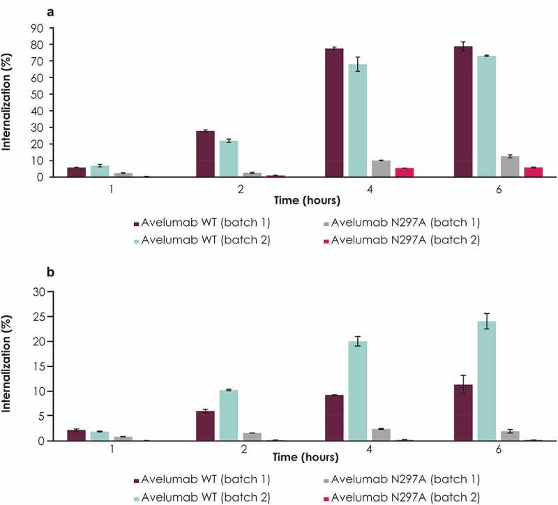
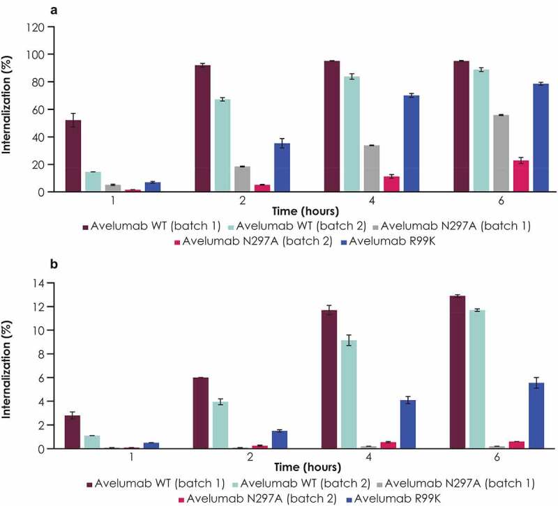
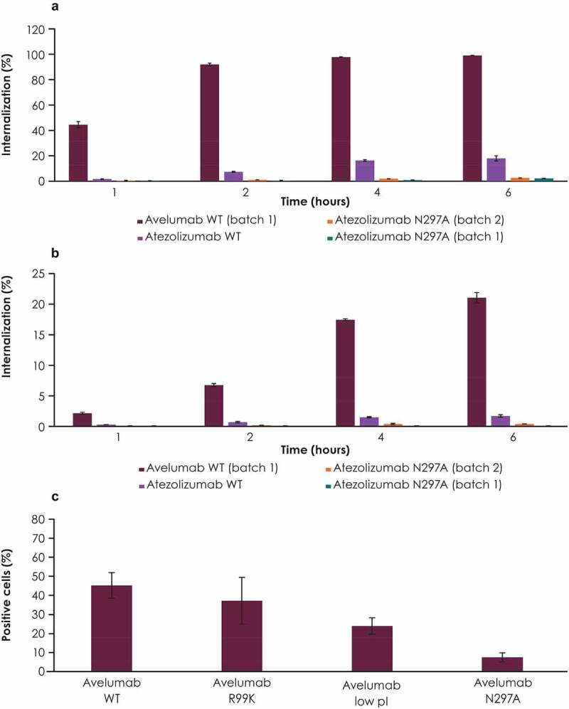
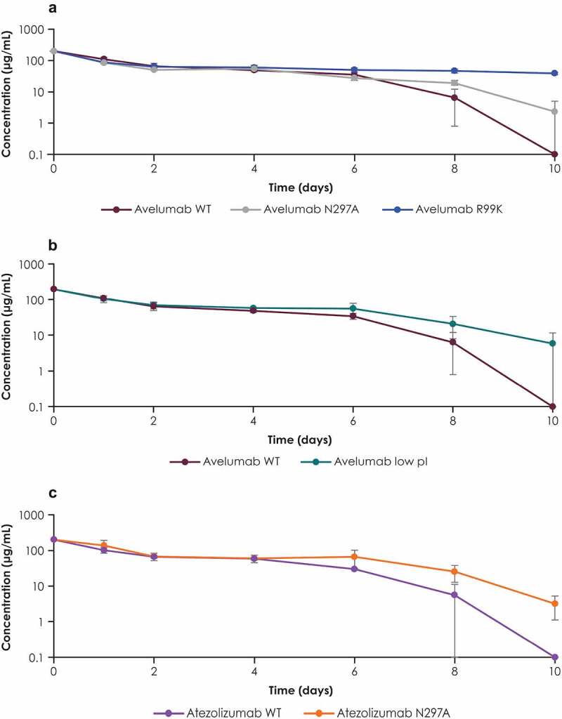
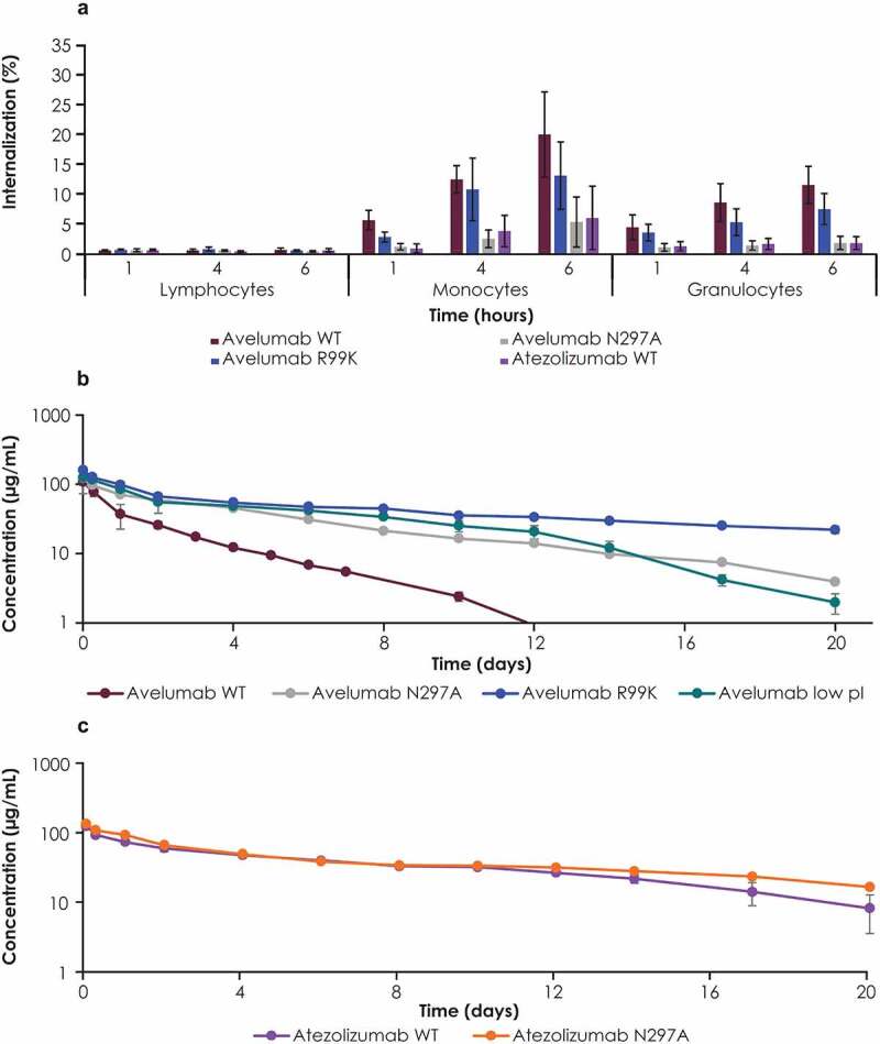
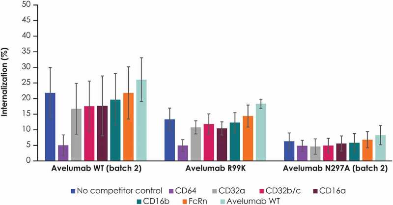
Avelumab is an IgG1 anti-programmed death ligand 1 (anti-PD-L1) monoclonal antibody that has been approved as a monotherapy for metastatic Merkel cell carcinoma and advanced urothelial carcinoma, and in combination with axitinib for advanced renal cell carcinoma. Avelumab is cleared faster and has a shorter half-life than other anti-PD-L1 antibodies, such as atezolizumab and durvalumab, but the mechanisms underlying these differences are unknown. IgG antibodies can be cleared through receptor-mediated endocytosis after binding of the antibody Fab region to target proteins, or via Fcγ receptor (FcγR)-mediated endocytosis. Unlike other approved anti-PD-L1 antibodies, avelumab has a native Fc region that retains FcγR binding capability. We hypothesized that the rapid clearance of avelumab might be due to the synergistic effect of both FcγR-mediated and PD-L1 target-mediated internalization. To investigate this, we performed in vitro and in vivo studies that compared engineered variants of avelumab and atezolizumab to determine mechanisms of cellular internalization. We found that both FcγR and PD-L1 binding contribute to avelumab internalization. While FcγR binding was the dominant mechanism of avelumab internalization in vitro, with CD64 acting as the most important FcγR, studies in mice and cynomolgus monkeys showed that both FcγR and PD-L1 contribute to avelumab elimination, with PD-L1 binding playing a greater role. These studies suggest that the rapid internalization of avelumab might be due to simultaneous binding of both PD-L1 and FcγR in trans. Our findings also provide a basis to alter the clearance and half-life of monoclonal antibodies in therapeutic development.

Keywords: Avelumab; FCγr binding; PD-L1 binding; clearance; internalization.


Figures

Figure 1.
Figure 1.
Internalization of WT avelumab compared with its FcγR binding–deficient variant (N297A) in human monocytes and granulocytes. Internalization in (a) CD14+ CD16− monocytes and (b) granulocytes. A flow cytometry–based antibody internalization assay with pH-sensitive fluorescent dye, pHrodo, was used to directly monitor antibody internalization in healthy donor blood. Blood samples were treated with pHrodo-labeled antibodies, and immune cells were detected with external staining antibodies. Error bars represent standard deviations. FcγR, Fcγ receptor; WT, wild type
Figure 2.
Figure 2.
Internalization of WT avelumab compared with its anti–PD-L1 binding–deficient variant (R99K) and FcγR binding–deficient variant (N297A) in total human monocytes and granulocytes. Internalization in (a) total monocytes and (b) granulocytes from a representative donor is shown. Results were similar in other donors (data not shown). Error bars represent standard deviations of replicate assays from the same donor. Anti–PD-L1, anti–programmed death ligand 1; WT, wild-type
Figure 3.
Figure 3.
Internalization of anti–PD-L1 antibodies compared with their variants. Internalization of WT avelumab was compared with antibodies containing the atezolizumab Fab domain attached to either an FcγR binding–deficient (N297A) or a WT IgG1 Fc domain (FcγR binding–restored) in (a) CD14+ CD16− monocytes and (b) granulocytes. (c) Internalization of WT avelumab compared with a low pI variant, as well as FcγR binding–deficient (N297A) and PD-L1 binding–deficient (R99K) variants, in human PBMCs. Error bars represent standard deviations. FcγR, Fcγ receptor; PBMC, peripheral blood mononuclear cell; PD-L1, programmed death ligand 1; pI, isoelectric point; WT, wild-type
Figure 4.
Figure 4.
Serum concentration profiles of PD-L1 antibodies and their variants in mice. Serum concentration profiles of (a) WT avelumab and its FcγR binding–deficient (N297A) and PD-L1 binding–deficient (R99K) variants, (b) WT avelumab and its low pI variant, and (c) WT atezolizumab and its FcγR binding–deficient variant (N297A). C57BL/6 mice were dosed with 200 µg of antibodies. Blood samples were collected at various time points, and serum concentrations were measured by immunoassay. Error bars represent standard deviations. FcγR, Fcγ receptor; PD-L1, programmed death ligand 1; pI, isoelectric point; WT, wild-type
Figure 5.
Figure 5.
Studies with PD-L1 antibodies and their variants in cynomolgus monkeys. (a) Internalization of avelumab and its FcγR binding–deficient (N297A) and PD-L1 binding–deficient (R99K) variants was assessed in whole blood samples from cynomolgus monkeys. Serum concentration profiles of (b) WT avelumab, n = 2; avelumab FcγR binding–deficient (N297A), n = 1; avelumab PD-L1 binding–deficient (R99K), n = 3; avelumab low pI: n = 3; and (c) WT atezolizumab with, n = 2 and atezolizumab N297A (FcγR binding-deficient), n = 1. Cynomolgus monkeys were dosed with 5 mg/kg of antibody variants (WT avelumab was dosed at 4 mg/kg and normalized to 5 mg/kg). Serum concentrations were measured by immunoassay; profiles affected by antidrug antibodies were excluded. Error bars represent standard deviations. FcγR, Fcγ receptor; pI, isoelectric point; WT, wild-type
Figure 6.
Figure 6.
Internalization of avelumab and its FcγR binding–deficient (N297K) and PD-L1 binding–deficient (R99K) variants in the presence of competing soluble receptors in humans blood samples. PBMCs were incubated with selected recombinant human receptor proteins or with WT avelumab, followed by selected fluorescently labeled antibodies. Internalization was assessed using external staining antibodies. Error bars represent standard deviations. PBMC, peripheral blood mononuclear cell; FcRN; neonatal Fc receptor; WT, wild-type

References

    1. Darvin P, Toor SM, Sasidharan Nair V, Elkord E.. Immune checkpoint inhibitors: recent progress and potential biomarkers. Exp Mol Med. 2018;50:1–10. - PMC - PubMed
    1. Hamilton G, Rath B. Avelumab: combining immune checkpoint inhibition and antibody-dependent cytotoxicity. Expert Opin Biol Ther. 2017;17:515–523. - PubMed
    1. Tang H, Liang Y, Anders RA, Taube JM, Qiu X, Mulgaonkar A, Liu X, Harrington SM, Guo J, Xin Y, et al. PD-L1 on host cells is essential for PD-L1 blockade-mediated tumor regression. J Clin Invest. 2018;128(2):580–588. - PMC - PubMed
    1. Lin H, Wei S, Hurt EM, Green MD, Zhao L, Vatan L, Szeliga W, Herbst R, Harms PW, Fecher LA, et al. Host expression of PD-L1 determines efficacy of PD-L1 pathway blockade-mediated tumor regression. J Clin Invest. 2018;128(4):1708. - PMC - PubMed
    1. Wu B, Sun X, Gupta HB, Yuan B, Li J, Ge F, Chiang H-C, Zhang X, Zhang C, Zhang D, et al. Adipose PD-L1 modulates PD-1/PD-L1 checkpoint blockade immunotherapy efficacy in breast cancer. Oncoimmunology. 2018;7(11):e1500107. - PMC - PubMed

Publication types