. 2021 Aug 16:11:646054.
doi: 10.3389/fcimb.2021.646054. eCollection 2021.

Microbial Warfare on Three Fronts: Mixed Biofilm of Aspergillus fumigatus and Staphylococcus aureus on Primary Cultures of Human Limbo-Corneal Fibroblasts

Affiliations

Microbial Warfare on Three Fronts: Mixed Biofilm of Aspergillus fumigatus and Staphylococcus aureus on Primary Cultures of Human Limbo-Corneal Fibroblasts

Adrián Ramírez-Granillo et al. Front Cell Infect Microbiol. .

Abstract

Background: Coinfections with fungi and bacteria in ocular pathologies are increasing at an alarming rate. Two of the main etiologic agents of infections on the corneal surface, such as Aspergillus fumigatus and Staphylococcus aureus, can form a biofilm. However, mixed fungal-bacterial biofilms are rarely reported in ocular infections. The implementation of cell cultures as a study model related to biofilm microbial keratitis will allow understanding the pathogenesis in the cornea. The cornea maintains a pathogen-free ocular surface in which human limbo-corneal fibroblast cells are part of its cell regeneration process. There are no reports of biofilm formation assays on limbo-corneal fibroblasts, as well as their behavior with a polymicrobial infection.

Objective: To determine the capacity of biofilm formation during this fungal-bacterial interaction on primary limbo-corneal fibroblast monolayers.

Results: The biofilm on the limbo-corneal fibroblast culture was analyzed by assessing biomass production and determining metabolic activity. Furthermore, the mixed biofilm effect on this cell culture was observed with several microscopy techniques. The single and mixed biofilm was higher on the limbo-corneal fibroblast monolayer than on abiotic surfaces. The A. fumigatus biofilm on the human limbo-corneal fibroblast culture showed a considerable decrease compared to the S. aureus biofilm on the limbo-corneal fibroblast monolayer. Moreover, the mixed biofilm had a lower density than that of the single biofilm. Antibiosis between A. fumigatus and S. aureus persisted during the challenge to limbo-corneal fibroblasts, but it seems that the fungus was more effectively inhibited.

Conclusion: This is the first report of mixed fungal-bacterial biofilm production and morphological characterization on the limbo-corneal fibroblast monolayer. Three antibiosis behaviors were observed between fungi, bacteria, and limbo-corneal fibroblasts. The mycophagy effect over A. fumigatus by S. aureus was exacerbated on the limbo-corneal fibroblast monolayer. During fungal-bacterial interactions, it appears that limbo-corneal fibroblasts showed some phagocytic activity, demonstrating tripartite relationships during coinfection.

Keywords: Aspergillus fumigatus; Staphylococcus aureus; fungal–bacterial interaction; human limbo-corneal fibroblast cells; mixed fungal–bacterial biofilms.


Conflict of interest statement

The authors declare that the research was conducted in the absence of any commercial or financial relationships that could be construed as a potential conflict of interest.

Figures

Figure 1
Figure 1
Quantification of microbial biofilms on primary HLFCs. Biomass quantification using the Christensen crystal violet method (CVM). AF (A), SA (D), and the FBI AF+SA (G) with and without HLFCs. Metabolic activity quantification using the tetrazolium salt reduction method (MTT) on the biofilm AF (B), SA (E), and AF+SA (H) with and without HLFCs. The results are four replicates of three different experiments: n = 12. Significance was determined using the Student–Newman–Keuls test with multicomparison of procedures and are indicated as: (*), p < 0.050. SEM micrographs of biofilms of AF+HLFC (C1: 1,000×, C2: 2,500×) and AF (C3: 1,000×, C4: 2,500×); biofilm of SA+HLFC (F1: 1,000×, F2: 5,000×) and SA (F3: 1,000×, F4: 5,000×); and biofilm of AF+SA+HLFC (I1: 1,000×, I2: 5,000×) and AF+SA (I3: 1,000×, I4: 5,000×). HLFCs, human limbo-corneal fibroblast cells; AF, Aspergillus fumigatus; SA, Staphylococcus aureus; H, hypha; F, fibroblast; A, anastomosis; Ch, channels; Asterisk (*), extracellular matrix; Eps, exopolymeric substance; ML, monolayer; Fp, filopodia; Cc, cocci.
Figure 2
Figure 2
MFBB on primary HLFCs. SEM micrographs of 24-h biofilm of AF+SA at different zooming (A: 1,000×; B: 5,000×; C: 10,000×). ECM air channels and fibroblast monolayer are observed (A). Cocci and hyphae are seen embedded in a condensed extracellular matrix (B, C). MFBB, mixed fungal–bacterial biofilm; HLFCs, human limbo-corneal fibroblast cells; AF, Aspergillus fumigatus; SA, Staphylococcus aureus; H, hypha; Ch, channels; Asterisk (*), extracellular matrix (ECM); ML, monolayer; Fp, filopodia; Cc, Cocci.
Figure 3
Figure 3
Biofilm formation on HLFCs at 24 h. HLFC: A monolayer by SEM [(A): 2,000×] and its ultrastructure by TEM (B: 6,000×; (C): 15,000×x). AF+HLFC: Fungal penetration.(D: 2,500×). Hyphae through the fibroblast (E: 7,500×; F: 20,000×). Fungal adhesion: Secretion of ECM over the HLFC (G: 2,500×; H: 2,500×; (I): 20,000×). SA+HLFC: The HLFCs form filopodia next to cocci (J: 10,000×). TEM showed cocci divided inside the cells. Note the formation of bacterial interstitial (K: 20,000×; (L): 40,000×). AF+SA+HLFC: SEM showed cocci affect the hyphae. HLFC forming filopodia. HLFC crescent form and filopodia next to cocci. Fungus appears destroyed (M: 10,000×). TEM showed the antibiosis effect over AF. Intracellular damage in the HLFC with the presence of vacuoles (N: 20,000×; O: 40,000×). HLFCs, human limbo-corneal fibroblast cells; AF, Aspergillus fumigatus; SA, Staphylococcus aureus; H, hypha; F, fibroblast; ML, monolayer; Fp, filopodia; asterisk (*), extracellular matrix; R, ribosomes; M, cytoplasmic membrane; Cg, collagen fibers; N, nucleus; Nc, nucleolus; V, transfer vesicles; G, secretion granules; Eps, exopolymeric substance; Cc, cocci; Bg, interstitial; Ph, phagocytosis.
Figure 4
Figure 4
Interaction between AF biofilm and HLFCs. SEM micrographs showed an antifungal effect caused by the HLFC at 24 h. The HLFC showed exosome-like structures that produce damage to the hyphae (A: 2,500×; B: 10,000×). Hypha damaged in the apical zone. (C: 2,500×; D: 10,000×). HLFCs, human limbo-corneal fibroblast cells; AF, Aspergillus fumigatus; H, hypha; F, fibroblast; A, anastomosis; asterisk (*), extracellular matrix; Ex, exosome like; ML, monolayer.
Figure 5
Figure 5
Molecular biofilm components detected on HLFCs. EFM micrograph at 24 h showed ECM composition over the HLFC monolayer. HLFC (A: 10×), AF (B: 10×), AF+HLFC (C: 10×), hyphal network and co-localization of carbohydrates and eDNA in the ECM. SA (D: 10×), SA+HLFC (E: 10×), co-localization of carbohydrates and eDNA in the center of the bacterial microcolonies growing over the fibroblasts. AF+SA (F: 40×) and AF+SA+HLFC (G: 40×) showed antibiosis against AF for both. Calcofluor White (CW: blue-chitin and glycosylated carbohydrates), concanavalin A (ConA: green-glucose and mannose residues), and propidium iodide (PI: red-nucleic acids). Co-localization was obtained by image merging. HLFCs, human limbo-corneal fibroblast cells; AF, Aspergillus fumigatus; SA, Staphylococcus aureus; H, hypha; F, fibroblast; Ch, channels; ML, monolayer; Cc, cocci; Co, co-localization; Mc, microcolony; N, nucleus.
Figure 6
Figure 6
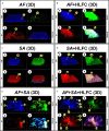
Construction of 3D models of the biofilms on HLFCs. AF (A): a biofilm rich in carbohydrates was detected. AF+HLFC (B): biofilm is denser with strong co-localization of the ECM components on the monolayer. In the bacterial 3D model, microcolonies are enveloped in layers of polysaccharides and eDNA, with stronger co-localization in SA+HLFC (D) compared with SA (C). The ECM was scarce for FBI models, AF+SA (E), and AF+SA+HLFC (F), detection of a strong antibiosis over AF. Calcofluor White (CW: blue-chitin and glycosylated carbohydrates), concanavalin A (ConA: green-glucose and mannose residues), and propidium iodide (PI: red-nucleic acids). Co-localization was obtained by image merging. HLFCs, human limbo-corneal fibroblast cells; AF, Aspergillus fumigatus; SA, Staphylococcus aureus; Ct, chitin; GM, N-acetylglucosamine/glucose and mannose residues; eD, extracellular DNA; F, fibroblast; Co, co-localization; H, hypha; Cc, cocci; N, nucleus.
Figure 7
Figure 7
Putative interaction of mixed infection on HLFCs related to eye infections. [MFBB] (A) Colonization of surface HLFCs by planktonic conidia. (B) A primary biofilm with rigid and abundant ECM, with planktonic propagules. Co-aggregation pathway could follow two different routes. (C) First pathway: secondary colonizer forms new bioscaffolds of sessile cocci. (D) Second pathway: planktonic bacteria induce co-aggregation over the fungal biofilm, reaching the sessile stage. Non-aggregate planktonic cells can migrate to another site. [ANTIBIOSIS] (E) Antagonistic effect from S. aureus over A. fumigatus. MICROORGANISMS AGAINST HLFC. (F) A. fumigatus causes hyphal perforation and EPS secretion, and hyphal adhesion. (G) S. aureus triggers pores and cellular lysis affecting membranes and cytoskeleton, breaking the desmosomes. HLFC AGAINST MICROORGANISMS. (H) HLFC against AF: Fibroblast produces exosomes and microvesicles that could be involved in phagocytic processes. (I) HLFC against SA: Fibroblast projects filopodia surrounding the cocci that are accompanied by modifications of the cytoskeleton (crescent formation). For details, see the Results section. Created with BioRender.com.

References

    1. Abelson M. B., McLaughlin J. (2012). Of Biomes, Biofilm and the Ocular Surface. Rev. Ophthalmol. 19 (9), 52.
    1. Allkja J., Bjarnsholt T., Coenye T., Cos P., Fallarero A., Harrison J. J., et al. . (2020). Minimum Information Guideline for Spectrophotometric and Fluorometric Methods to Assess Biofilm Formation in Microplates. Biofilm. 2, 100010. 10.1016/j.bioflm.2019.100010 - DOI - PMC - PubMed
    1. Álvarez-Félix J. R., Palazuelos-Gaxiola M., López-López G. (2010). Prevalencia De Queratitis En El Servicio De Oftalmología De La Coordinación Universitaria Del Hospital Civil De Culiacán. Rev. Med. UAS Nueva época 1 (3), 11–14.
    1. Archer N. K., Mazaitis M. J., Costerton J. W., Leid J. G., Powers M. E., Shirtliff M. E. (2011). Staphylococcus Aureus Biofilms: Properties, Regulation and Roles in Human Disease. Virulence 2 (5), 445–459. 10.4161/viru.2.5.17724 - DOI - PMC - PubMed
    1. Arvanitis M., Mylonakis E. (2015). Fungal–bacterial Interactions and Their Relevance in Health. Cell. Microbiol. 17 (10), 1442–1446. 10.1111/cmi.12493 - DOI - PubMed

Publication types

LinkOut - more resources