

Unilateral retinopathy post perilesional interferon α2b injections for ocular surface squamous cell carcinoma
- PMID: 34485759
- PMCID: PMC8405888
- DOI: 10.1016/j.ajoc.2021.101196
Unilateral retinopathy post perilesional interferon α2b injections for ocular surface squamous cell carcinoma
Abstract
Purpose: To describe the clinical course of a patient presenting with unilateral retinopathy after perilesional interferon alpha injections for treatment of ocular surface squamous cell carcinoma.
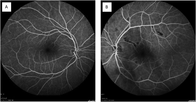
Observations: A patient, who was being treated with interferon alpha for ocular squamous cell carcinoma, presented with new onset decreased vision in her left eye. Upon examination, she was found to have cotton wool spots and retinal hemorrhages in the affected eye.
Conclusions and importance: Retinopathy is a well-documented side effect of systemic usage of interferon alpha. However, retinopathy has not been well discussed in the scenario of perilesional injections of interferon. It is important for clinicians to monitor for such pathology when using interferon alpha not only systemically, but also locally.
Keywords: Interferon α2b; Ocular squamous neoplasia; Retinopathy.
© 2021 The Authors.
Figures


References
-
- Rentiya Z.S., Wells M., Bae J., et al. Interferon-α-induced retinopathy in chronic hepatitis C treatment: summary, considerations, and recommendations. Graefes Arch Clin Exp Ophthalmol. 2019;257:447–452. - PubMed
-
- Raza A., Mittal S., Sood G.K. Interferon-associated retinopathy during the treatment of chronic hepatitis C: a systematic review. J Viral Hepat. 2013;20:593–599. - PubMed
-
- Vann R.R., Karp C.L. Perilesional and topical interferon alfa-2b for conjunctival and corneal neoplasia. Ophthalmology. 1999;106:91–97. - PubMed
-
- Manhard J.C., Mary . 2018. Unilateral Interferon-Associated Retinopathy After Repeated Perilesional Interferon α2b Injections for Ocular Surface Squamous Cell Carcinoma. ASRS Annual Meeting. Vancouver, BC, Canada.
Publication types
LinkOut - more resources
Full Text Sources
