. 2021 Jul;5(4):441-452.
doi: 10.1109/trpms.2020.3020212. Epub 2020 Aug 28.

Photon Counting CT: Clinical Applications and Future Developments

Affiliations

Photon Counting CT: Clinical Applications and Future Developments

Scott S Hsieh et al. IEEE Trans Radiat Plasma Med Sci. 2021 Jul.

Abstract

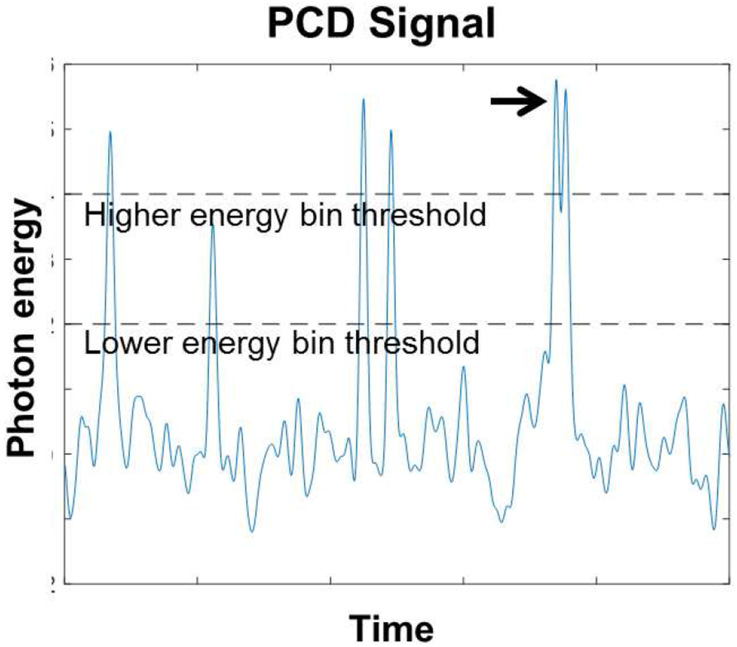
The use of a photon counting detector in CT (PCD CT) is currently the subject of intense investigation and development. In this review article, we will describe potential clinical applications of this technology with a particular focus on the experience of our own institution with a prototype PCD CT scanner. PCDs have three primary advantages over conventional, energy integrating detectors (EIDs): they provide spectral information without need for a dedicated dual energy protocol; they are immune to electronic noise; and they can be made very high resolution without significant compromises to quantum efficiency. These advantages translate into several clinical applications. Metal artifacts, beam hardening artifacts, and noise streaks from photon starvation can be better mitigated using PCD CT. Certain incidental findings can be better characterized using the spectral information from PCD CT. High-contrast, high-resolution structures such as the temporal bone can be better visualized using PCD CT and at greatly reduced dose. We also discuss new possibilities on the horizon, including new contrast agents, and how anticipated improvements in PCD CT will translate to performance in these applications.

Keywords: clinical applications; photon counting X-ray detectors; spectral CT.


Figures

Fig. 1.
Fig. 1.
Simulated example of a signal detected by a PCD. Impinging photons deposit energy in the PCD, which creates a momentary signal with amplitude ideally proportional to the deposited energy. Six photons are present in this example, each one leading to a peak. If the amplitude exceeds preset comparator thresholds (dashed lines), the PCD increments the corresponding counter. The black arrow points to an example of pileup, wherein two photons arrive in close succession. In this case, the low energy bin is incremented only once even though two photons have arrived. Pileup and charge sharing (not shown here) are two major sources of error in PCDs. If the lower energy bin threshold is set sufficiently high, it will almost never be triggered from electronic noise (which appear as random fluctuations in this plot). Hence, the number of counts stemming from electronic noise can be close to zero in practice.
Fig. 2.
Fig. 2.
Improvement in CNR using a PCD, demonstrated in an anthropomorphic phantom. (Left) EID CT at 140 kVp. (Right) PCD CT with energy bin thresholds at 25 and 65 keV, scanned using the same protocol. The iodine contrast is improved. Adapted from Reference [4] with permission.
Fig. 3.
Fig. 3.
Anthropomorphic phantom showing improvement in metal artifacts from (left) EID CT to (right) high-threshold PCD CT with tin filtration. (WW, WL) = (400, 40) HU. Adapted from Reference [1] with permission.
Fig. 4.
Fig. 4.
Scan of an anthropomorphic phantom at 20 mAs with (left) EID and (right) PCD CT. The EID CT shows noise streaks and photon starvation near the center of the object. Adapted from Reference [2] with permission.
Fig. 5.
Fig. 5.
Comparison of dose efficiency for very high resolution tasks. (Top and center rows) Cadaver scan of a pelvis, comparing an energy-integrating detector (EI) with a PCD operating in either macro mode or UHR mode, with the UHR mode using smaller pixel size. All systems were scanned at the same dose. UHR mode shows improved noise using the B70f kernel. Adapted from [5] with permission. (Bottom row) The left and right columns shows EID and PCD CT, respectively. PCD CT reduces dose by (top row) 85% and (bottom row) 81% without reducing high-resolution detectability. The EID CT uses a comb filter, which reduces dose efficiency. (WW, WL) = (3200, 700) HU. Adapted from Reference [9] with permission.
Fig. 6.
Fig. 6.
Detection of kidney stones with (left) EID and (right) PCD CT. The improved resolution of PCD enables better detectability for these small, high contrast objects. The enhanced spectral capabilities also enable better classification of the stone type. Adapted from Reference [3].
Fig. 7.
Fig. 7.
Comparison of a Promus Premier stent placed in a rabbit model, scanned using either a conventional EID CT scanner (Philips Brilliance 64) or with a prototype spectral photon counting CT (SPCCT) scanner, which is also capable of providing a platinum-only reconstruction. The details of the stent can be more cleanly visualized with SPCCT. Adapted from Reference [6] with permission.
Fig. 8.
Fig. 8.
Volumetric rendering of an extremity PCD CT reconstruction of a human volunteer wrist including a wristwatch, scanned with the prototype MARS scanner. Image courtesy of MARS Bioimaging. Adapted from [RK Panta, IEEE 2018]. (©2018 IEEE).

References

    1. Zhou W et al. , “Reduction of metal artifacts and improvement in dose efficiency using photon-counting detector computed tomography and tin filtration,” Investigative radiology, vol. 54, no. 4, pp. 204–211, 2019. - PMC - PubMed
    1. Yu Z et al. , “Noise performance of low-dose CT: comparison between an energy integrating detector and a photon counting detector using a whole-body research photon counting CT scanner,” Journal of Medical Imaging, vol. 3, no. 4, p. 043503, 2016. - PMC - PubMed
    1. Marcus RP et al. , “Detection and Characterization of Renal Stones by Using Photon-Counting–based CT,” Radiology, vol. 289, no. 2, pp. 436–442, 2018. - PMC - PubMed
    1. Yu Z et al. , “Evaluation of conventional imaging performance in a research whole-body CT system with a photon-counting detector array,” Physics in Medicine & Biology, vol. 61, no. 4, p. 1572, 2016. - PMC - PubMed
    1. Klein L et al. , “Effects of Detector Sampling on Noise Reduction in Clinical Photon-Counting Whole-Body Computed Tomography,” Investigative radiology, vol. 55, no. 2, pp. 111–119, 2020. - PubMed

LinkOut - more resources