Review
. 2023 Jun;64(3):262-283.
doi: 10.1007/s12016-021-08889-8. Epub 2021 Sep 6.

Current Concepts on the Pathogenesis of Systemic Sclerosis

Affiliations
Review

Current Concepts on the Pathogenesis of Systemic Sclerosis

Marie Elise Truchetet et al. Clin Rev Allergy Immunol. 2023 Jun.

Abstract

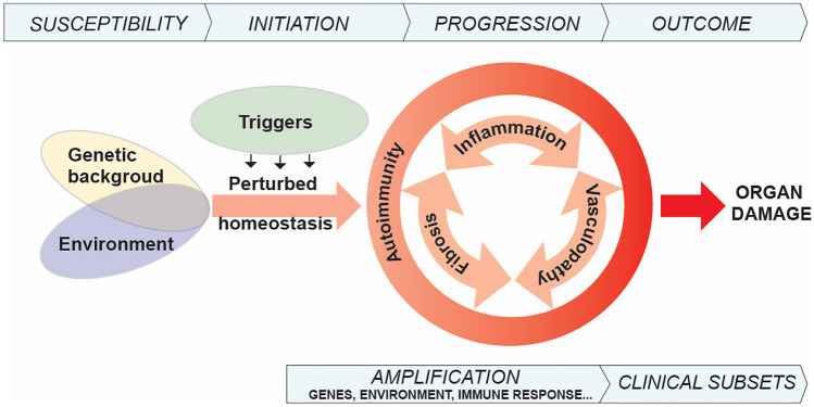
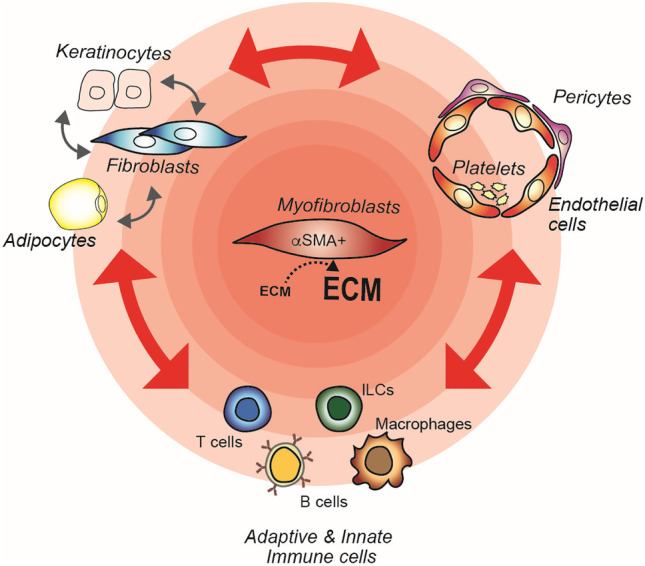
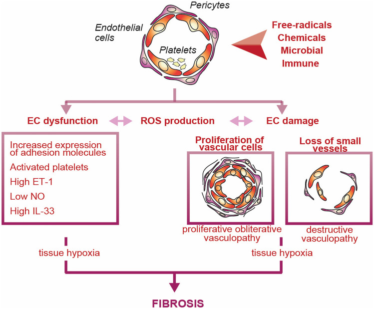
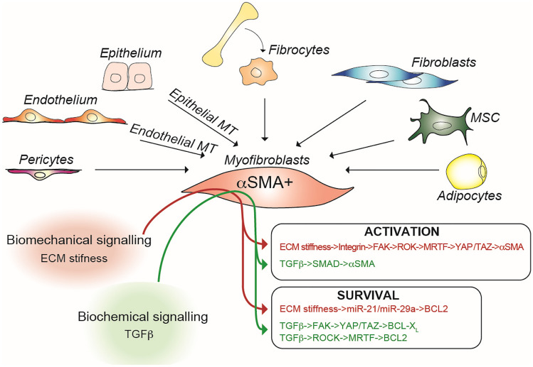
From the clinical standpoint, systemic sclerosis (SSc) is characterized by skin and internal organ fibrosis, diffuse fibroproliferative vascular modifications, and autoimmunity. Clinical presentation and course are highly heterogenous and life expectancy variably affected mostly dependent on lung and heart involvement. SSc touches more women than men with differences in disease severity and environmental exposure. Pathogenetic events originate from altered homeostasis favored by genetic predisposition, environmental cues and a variety of endogenous and exogenous triggers. Epigenetic modifications modulate SSc pathogenesis which strikingly associate profound immune-inflammatory dysregulation, abnormal endothelial cell behavior, and cell trans-differentiation into myofibroblasts. SSc myofibroblasts show enhanced survival and enhanced extracellular matrix deposition presenting altered structure and altered physicochemical properties. Additional cell types of likely pathogenic importance are pericytes, platelets, and keratinocytes in conjunction with their relationship with vessel wall cells and fibroblasts. In SSc, the profibrotic milieu is favored by cell signaling initiated in the one hand by transforming growth factor-beta and related cytokines and in the other hand by innate and adaptive type 2 immune responses. Radical oxygen species and invariant receptors sensing danger participate to altered cell behavior. Conventional and SSc-specific T cell subsets modulate both fibroblasts as well as endothelial cell dysfunction. Beside autoantibodies directed against ubiquitous antigens important for enhanced clinical classification, antigen-specific agonistic autoantibodies may have a pathogenic role. Recent studies based on single-cell RNAseq and multi-omics approaches are revealing unforeseen heterogeneity in SSc cell differentiation and functional states. Advances in system biology applied to the wealth of data generated by unbiased screening are allowing to subgroup patients based on distinct pathogenic mechanisms. Deciphering heterogeneity in pathogenic mechanisms will pave the way to highly needed personalized therapeutic approaches.

Keywords: Fibrosis; Immune responses; Inflammation; Pathogenesis; System biology; Systemic sclerosis.


Conflict of interest statement

The authors declare no competing interests.

Figures

Fig. 1
Fig. 1
Overview of conditions and events leading to systemic sclerosis. Schematic diagram highlighting the complex interplay thought to play a role in susceptibility and initiation of SSc in which genetic predisposition and environmental cues under the pressure of a variety of triggers lead to perturbed homeostasis with ensuing autoimmunity. Autoimmunity is represented as the common denominator of the three fundamental aspects of SSc: inflammation, vasculopathy, and fibrosis. Heterogeneous clinical manifestations would then develop according to variable amplification mechanisms resulting in recognized clinical subsets and organ damage
Fig. 2
Fig. 2
Major cell types and their multiple interactions in SSc pathogenesis. SSc is here viewed as a three-leg pathology in which major dysfunctional cell types are immune cells, endothelial cells, and fibroblasts which directly or indirectly intensely interact leading to myofibroblast hyperactivation. This cell and soluble factor three-leg network establishes further interactions with many other cell types including adipocytes, keratinocytes, pericytes, and platelets. The concentric reddish shadow highlights the influence of the various cell types on the activation of myofibroblasts. Two-head red arrows indicate multiple, reciprocal interactions mainly ensured by soluble mediators of inflammation. Dashed arrow indicates increase in extracellular matrix (ECM) deposition by myofibroblasts
Fig. 3
Fig. 3
Vasculopathy in SSc. In SSc, under the influence of a variety of stimuli here depicted as a bicolor arrowhead, EC become dysfunctional and undergo damage. Excess in vasoconstricting over vasodilating agents, as well as enhanced fibroproliferative events of the vessel wall associated with reduced angiogenesis and vasculogenesis are characteristic. Vasculopathic alterations contribute to the developments of fibrosis. ET-1, endothelin-1; IL, interleukin; NO, nitric oxide; ROS, radical oxygen species
Fig. 4
Fig. 4
Myofibroblasts and their centrality in the development of fibrosis in SSc. Depicted are the cells potentially giving origin to myofibroblasts, as well as the main signals involved in their activation and survival. αSMA, alpha smooth muscle actin; BCL, B-cell lymphoma; ECM, extracellular matrix; FAK, focal adhesion kinase; MRTF, myocardin-related transcription factors; ROCK, Rho-associated creatinine kinase; SMAD, small mothers against decapentaplegic; TAZ, transcriptional co-activator with PDZ-binding motif; TGF-β, transforming growth factor-beta; YAP, Yes kinase-associated protein
Fig. 5
Fig. 5
Altered cross-talk between keratinocytes and dermal fibroblasts in SSc. The homeostatic relationship between epidermis and dermis includes reciprocal signaling here represented by IL-1 produced by keratinocytes and KGF by fibroblasts. Cytokines dysregulated in SSc alter this cross-talk and variably affect the inflammatory and ECM deposition properties of dermal fibroblasts. ECM, extracellular matrix; KGF, keratinocyte growth factor; IL, interleukin; TGF-β, transforming growth factor beta; TNF, tumor necrosis factor
Fig. 6
Fig. 6
Contribution of cells and soluble products of the innate immune system to enhanced ECM deposition. Parallel, not mutually exclusive pathways involving cells of the innate immune system and their soluble products, converge on fibroblasts enhancing their ECM synthetic capacity. CXCL, chemokine containing the CXC motif; DAMP, danger associated molecular patterns; IFN-I, type I interferon; IL, interleukin; ILC, innate lymphoid cell; pDC, plasmacytoid dendritic cell; TGF-β, transforming growth factor-beta; Th2, type 2 T helper cell; TNF, tumor necrosis factor; TSLP, thymic stromal lympopoietin
Fig. 7
Fig. 7
Adaptive immune responses and their roles in SSc. T cells, B cells, and their products contribute to both enhanced ECM deposition and vasculopathy. This schematic representation highlights the characteristics of conventionally defined as well as of SSc-restricted T cell subsets. They may have enhancing or inhibitory functions (blunted heads: inhibitory function; arrowhead: enhancing function). T-B cell interactions are important for both the generation of agonist/antagonist autoAb and tissue damage. CD, cluster of differentiation; CXCL, chemokine containing the CXC motif; IFN-γ, interferon-gamma; IL, interleukin; TFH, T follicular helper cell; Th, T helper cell. Autoantibody specificities: AFA, anti-fibroblast; AT1R, angiotensin-II receptor; ECA, endothelial cell; ETAR, endothein-1 receptor A; MMP, matrix metalloproteinase; NAG2, also known as transmembrane 4 superfamily member 7; PDGF-Rα, platelet-derived growth factor receptor-alpha; UL-94, gene coding for the cytomegalovirus (CMV) cytoplasmic envelopment protein 2

References

    1. Denton CP, Khanna D. Systemic sclerosis. Lancet. 2017;390:1685–1699. doi: 10.1016/s0140-6736(17)30933-9. - DOI - PubMed
    1. Elhai M, Meune C, Boubaya M, Avouac J, Hachulla E, Balbir-Gurman A, Riemekasten G, Airo P, Joven B, Vettori S, Cozzi F, Ullman S, Czirjak L, Tikly M, Muller-Ladner U, Caramaschi P, Distler O, Iannone F, Ananieva LP, Hesselstrand R, Becvar R, Gabrielli A, Damjanov N, Salvador MJ, Riccieri V, Mihai C, Szucs G, Walker UA, Hunzelmann N, Martinovic D, Smith V, Muller CS, Montecucco CM, Opris D, Ingegnoli F, Vlachoyiannopoulos PG, Stamenkovic B, Rosato E, Heitmann S, Distler JHW, Zenone T, Seidel M, Vacca A, Langhe E, Novak S, Cutolo M, Mouthon L, Henes J, Chizzolini C, Muhlen CAV, Solanki K, Rednic S, Stamp L, Anic B, Santamaria VO, De Santis M, Yavuz S, Sifuentes-Giraldo WA, Chatelus E, Stork J, Laar JV, Loyo E, de la Pena Garcia, Lefebvre P, Eyerich K, Cosentino V, Alegre-Sancho JJ, Kowal-Bielecka O, Rey G, Matucci-Cerinic M, Allanore Y. Mapping and predicting mortality from systemic sclerosis. Ann Rheum Dis. 2017;76:1897–1905. doi: 10.1136/annrheumdis-2017-211448. - DOI - PubMed
    1. Moore DF, Steen VD. Racial disparities in systemic sclerosis. Rheum Dis Clin North Am. 2020;46:705–712. doi: 10.1016/j.rdc.2020.07.009. - DOI - PubMed
    1. Abraham DJ, Varga J. Scleroderma: from cell and molecular mechanisms to disease models. Trends Immunol. 2005;26:587–595. doi: 10.1016/j.it.2005.09.004. - DOI - PubMed
    1. Katsumoto TR, Whitfield ML, Connolly MK. The pathogenesis of systemic sclerosis. Annual review of pathology. 2011;6:509–537. doi: 10.1146/annurev-pathol-011110-130312. - DOI - PubMed