. 2022 Jan;123(1):128-141.
doi: 10.1002/jcb.30140. Epub 2021 Sep 6.

Pharmacological inhibition of BAG3-HSP70 with the proposed cancer therapeutic JG-98 is toxic for cardiomyocytes

Affiliations

Pharmacological inhibition of BAG3-HSP70 with the proposed cancer therapeutic JG-98 is toxic for cardiomyocytes

Thomas G Martin et al. J Cell Biochem. 2022 Jan.

Abstract

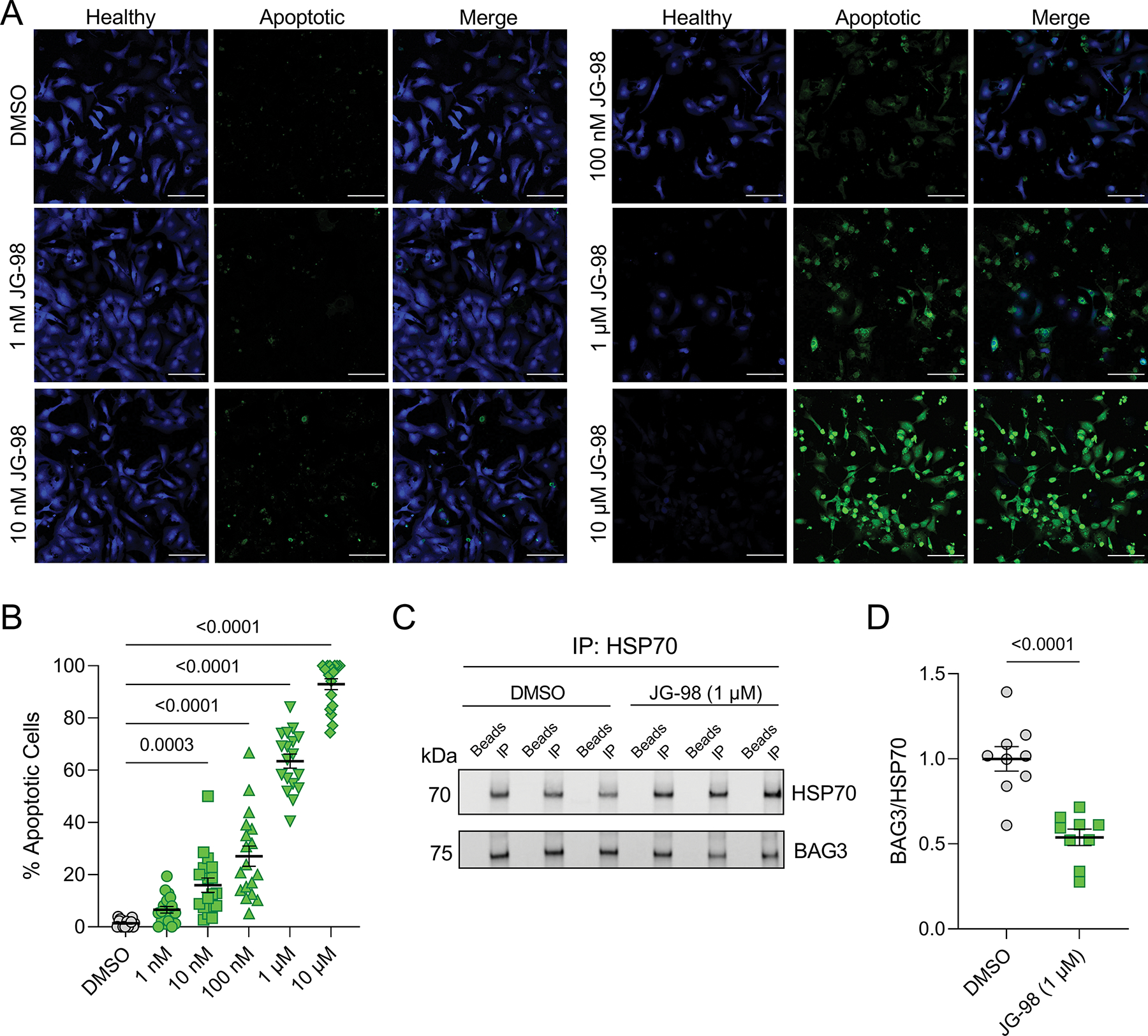
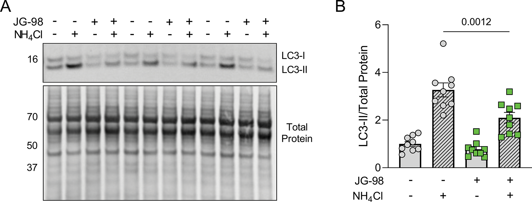
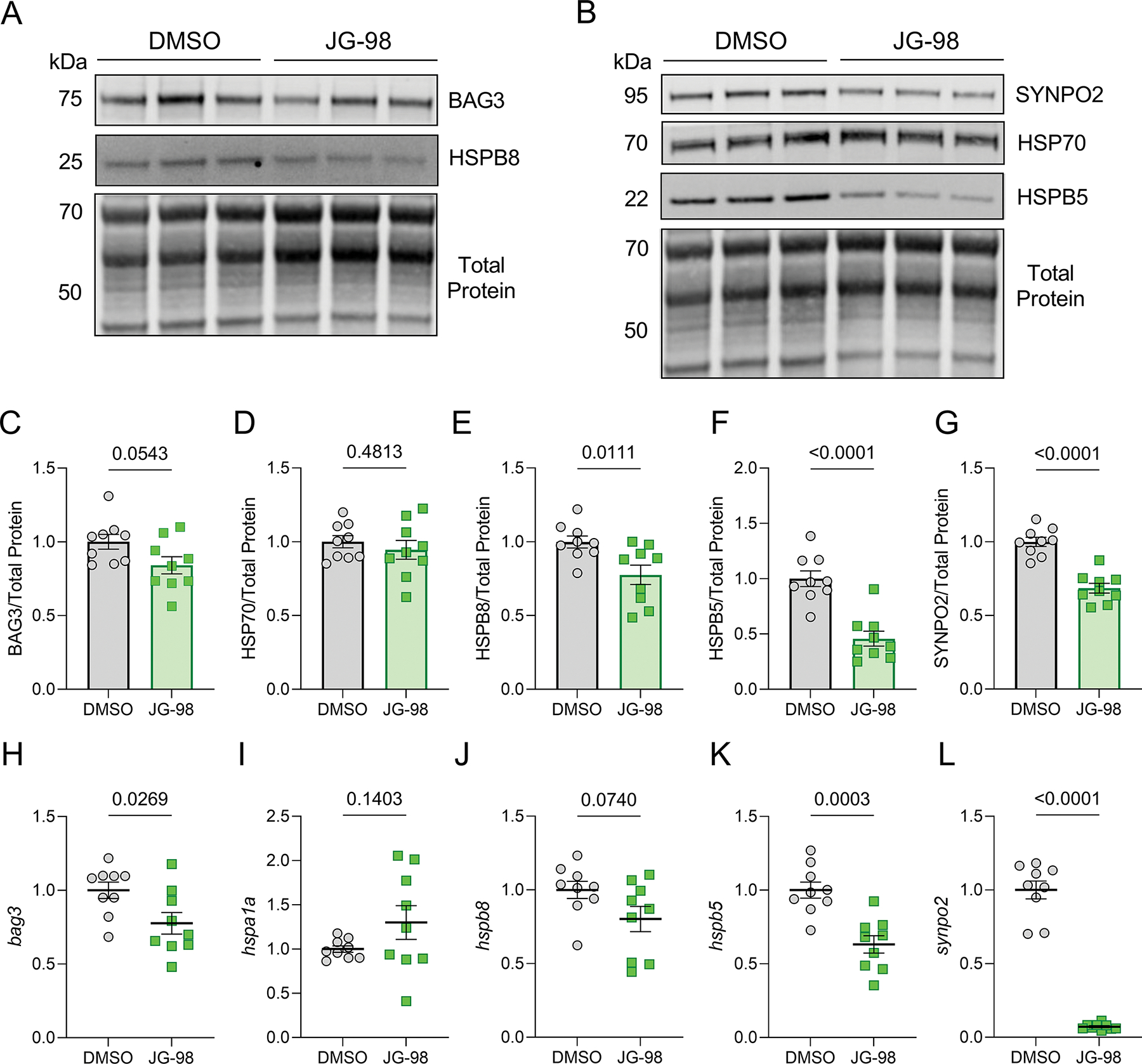
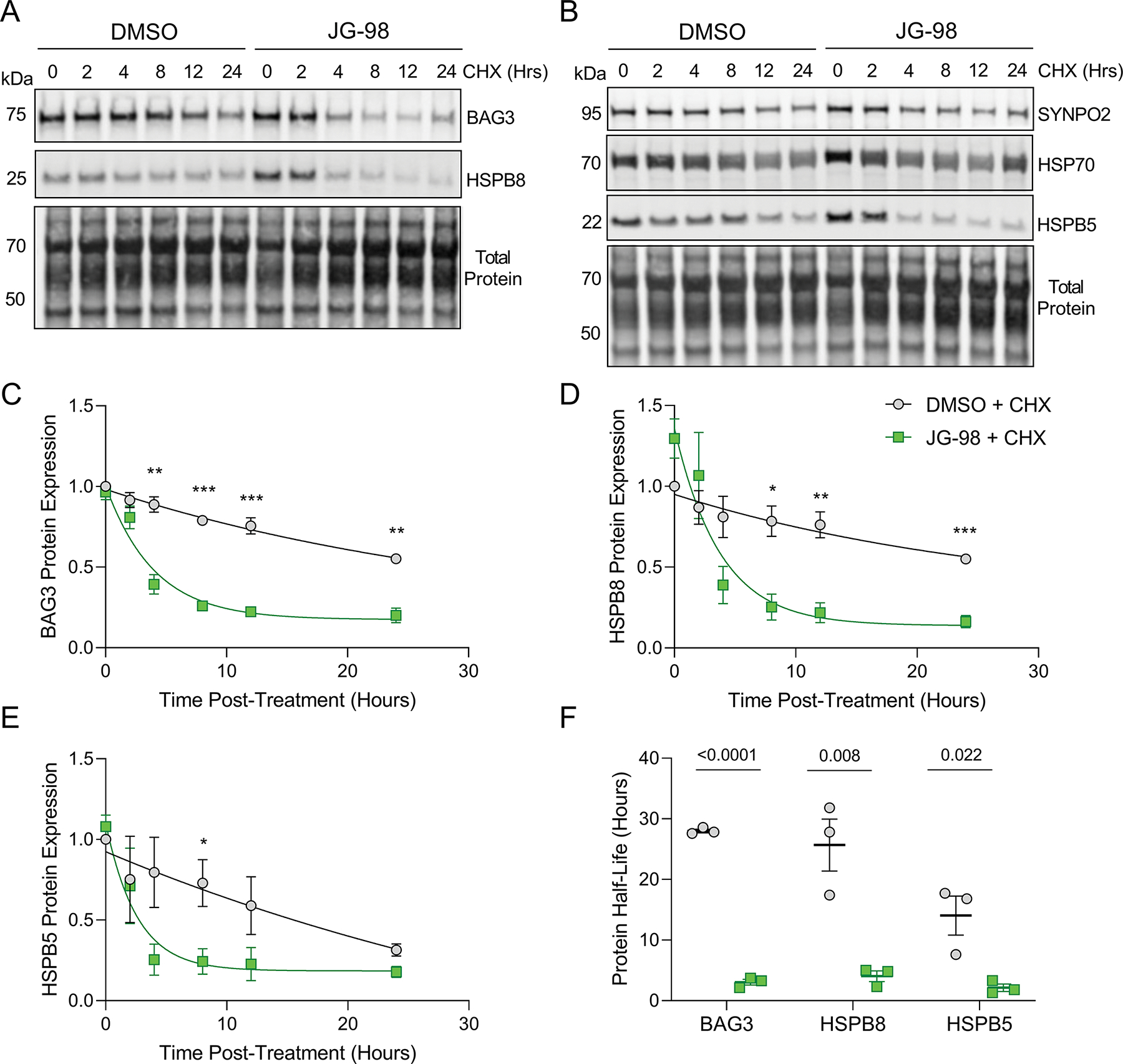
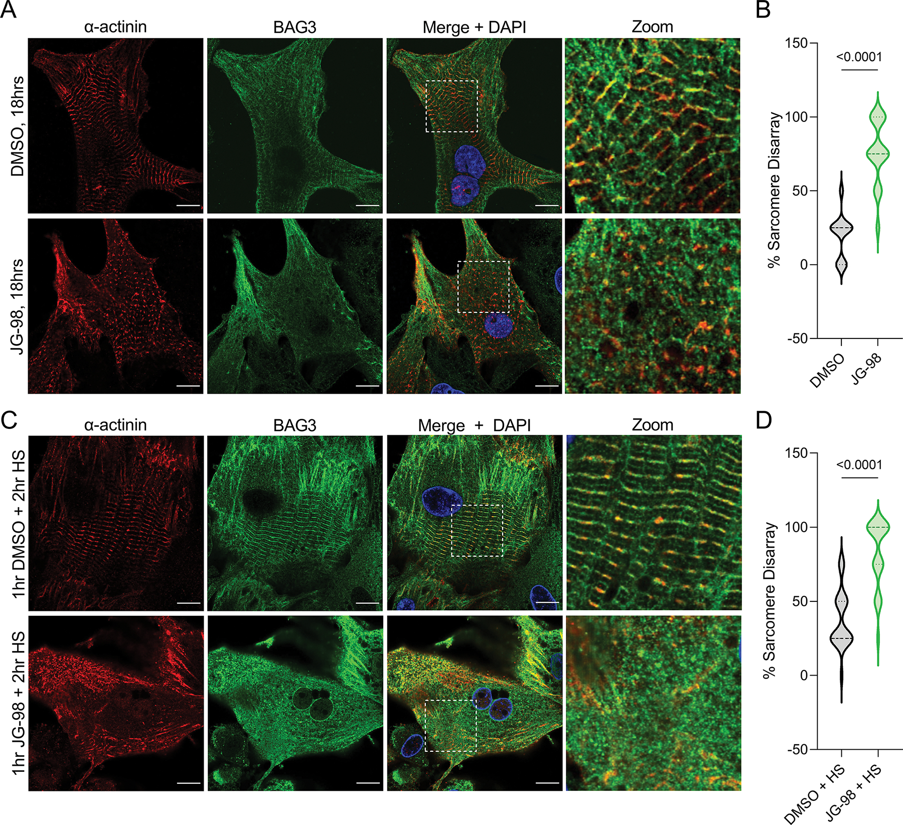
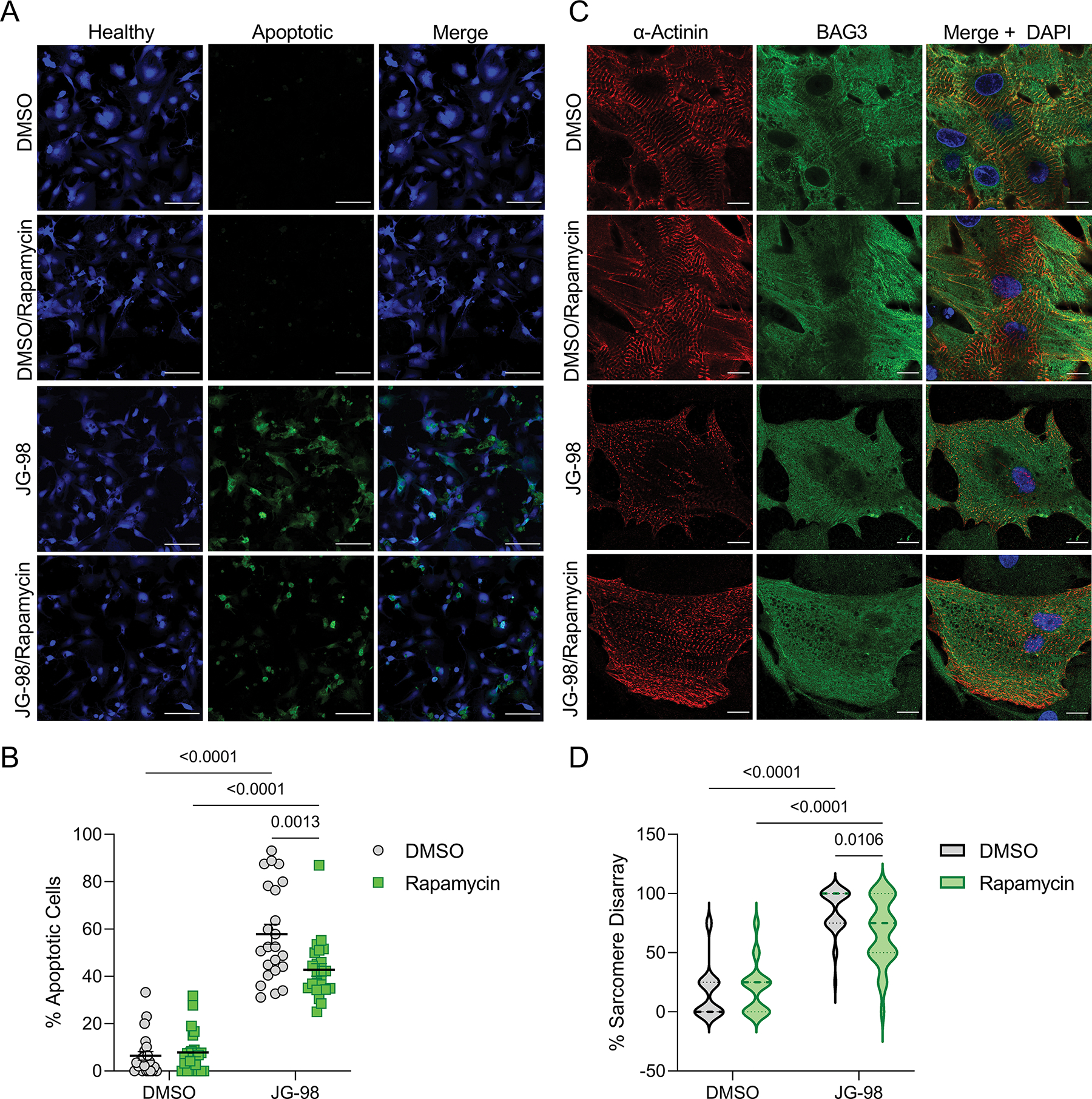
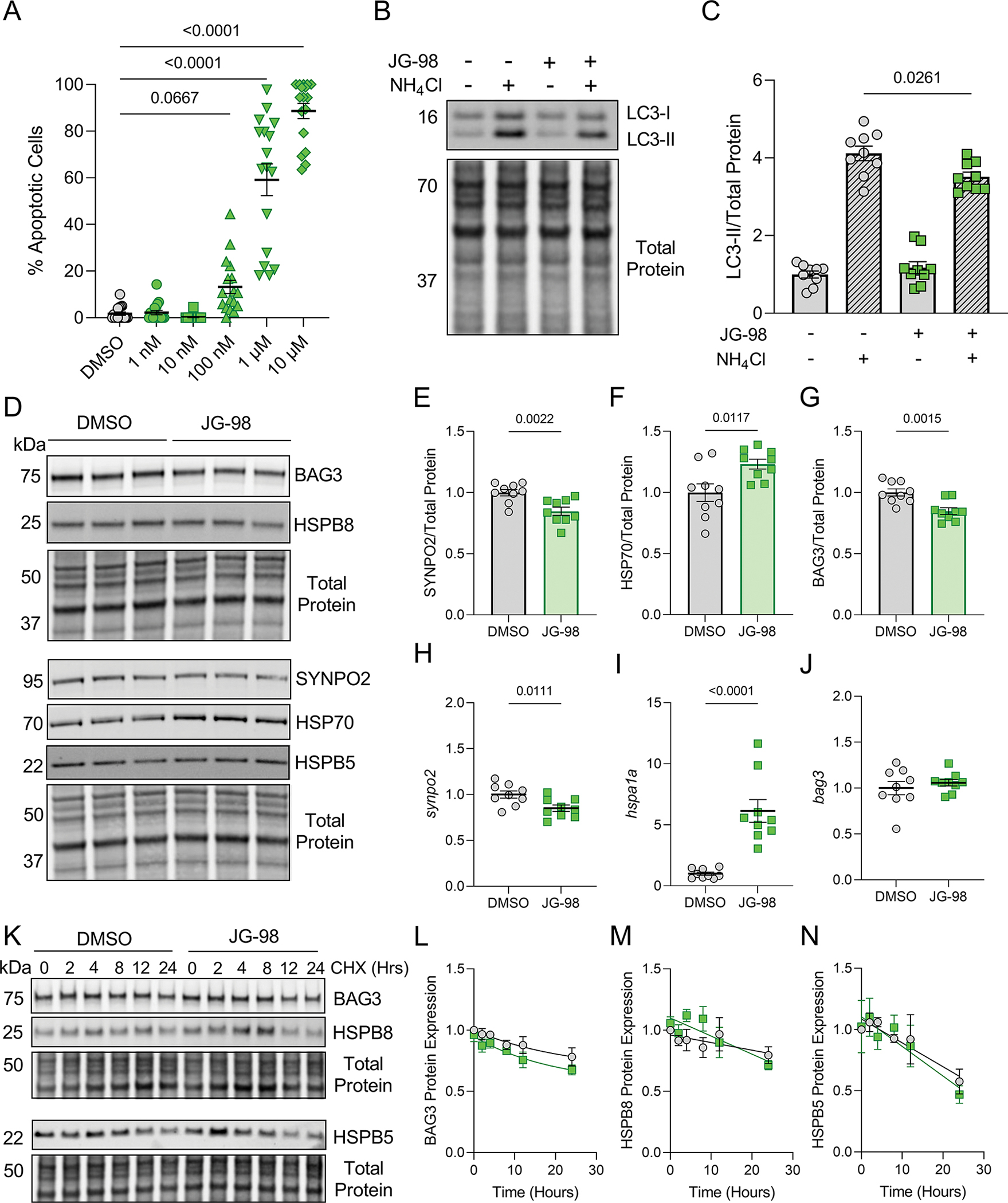
The co-chaperone Bcl2-associated athanogene-3 (BAG3) maintains cellular protein quality control through the regulation of heat shock protein 70 (HSP70). Cancer cells manipulate BAG3-HSP70-regulated pathways for tumor initiation and proliferation, which has led to the development of promising small molecule therapies, such as JG-98, which inhibit the BAG3-HSP70 interaction and mitigate tumor growth. However, it is not known how these broad therapies impact cardiomyocytes, where the BAG3-HSP70 complex is a key regulator of protein turnover and contractility. Here, we show that JG-98 exposure is toxic in neonatal rat ventricular myocytes (NRVMs). Using immunofluorescence microscopy to assess cell death, we found that apoptosis increased in NRVMs treated with JG-98 doses as low as 10 nM. JG-98 treatment also reduced autophagy flux and altered expression of BAG3 and several binding partners involved in BAG3-dependent autophagy, including SYNPO2 and HSPB8. We next assessed protein half-life with disruption of the BAG3-HSP70 complex by treating with JG-98 in the presence of cycloheximide and found BAG3, HSPB5, and HSPB8 half-lives were reduced, indicating that complex formation with HSP70 is important for their stability. Next, we assessed sarcomere structure using super-resolution microscopy and found that disrupting the interaction with HSP70 leads to sarcomere structural disintegration. To determine whether the effects of JG-98 could be mitigated by pharmacological autophagy induction, we cotreated NRVMs with rapamycin, which partially reduced the extent of apoptosis and sarcomere disarray. Finally, we investigated whether the effects of JG-98 extended to skeletal myocytes using C2C12 myotubes and found again increased apoptosis and reduced autophagic flux. Together, our data suggest that nonspecific targeting of the BAG3-HSP70 complex to treat cancer may be detrimental for cardiac and skeletal myocytes.

Keywords: BAG3; HSP70; JG-98; cancer therapy; cardio-oncology; cardiomyocyte; cytotoxicity.


Conflict of interest statement

CONFLICTS OF INTEREST

The authors declare that there are no conflicts of interest.

Figures

Figure 1.
Figure 1.. Disrupting the BAG3-HSP70 complex with JG-98 causes apoptosis in cardiomyocytes.
A, Representative immunofluorescence images for NRVMs treated with DMSO and 1 nM to 10 μM JG-98; green – extracellular membrane phosphatidylserine (apoptotic), blue – healthy; scale bars = 100 μm. B, Quantification of apoptosis in the immunofluorescence images; n = 18 images per treatment from 3 separate experiments; data were analyzed by one-way ANOVA with Tukey’s post-hoc test. C, Representative western blot for HSP70 and BAG3 in HSP70 immunoprecipitation experiments from cells treated with DMSO or 1 μM JG-98. D, Quantitative densitometry analysis of the BAG3 signal intensity normalized to HSP70; n = 9 per group from 3 separate experiments; data were analyzed by two-tailed t-test. All data are presented as the mean ± SEM.
Figure 2.
Figure 2.. JG-98 treatment reduces autophagy flux.
A, Representative western blot for LC3 in NRVMs treated with DMSO or JG-98 in the presence or absence of the autophagy inhibitor NH4Cl. B, Quantification of LC3-II expression normalized to total protein; n = 9 per group from 3 separate experiments; data were analyzed with two-way ANOVA (interaction, p = 0.023) with Tukey’s post-hoc test. Data are presented as the mean ± SEM.
Figure 3.
Figure 3.. Inhibition of BAG3-HSP70 alters expression of proteins involved in BAG3-dependent autophagy pathway.
A-B, Representative western blots for BAG3, HSPB8, SYNPO2, HSP70, HSPB5 in NRVMs treated with DMSO or JG-98. C-G, Quantification of BAG3 (C), HSP70 (D), HSPB8 (E), HSPB5 (F), and SYNPO2 (G) protein expression normalized to total protein loading control. H-L, qPCR analysis of bag3 (H), hspa1a (I), hspb8 (J), hspb5 (K), and synpo2 (L) mRNA expression normalized to β-actin. All treatments were with 1 μM JG-98 for 18 hours. For all, n = 9 per group from 3 separate experiments; data were analyzed by two-tailed t-test and are presented as the mean ± SEM.
Figure 4.
Figure 4.. The BAG3-HSP70 interaction is required for maintaining BAG3, HSPB8, and HSPB5 protein stability.
A-B, Representative western blots for BAG3, HSPB8, SYNPO2, HSP70, HSPB5 in NRVMs treated with DMSO or 1 μM JG-98 in the presence of cycloheximide (CHX) for 0–24 hours. C-E, Quantification of BAG3 (C), HSPB8 (D), and HSPB5 (E) protein expression normalized to total protein over the time-course of CHX treatment with DMSO or JG-98; n = 3 for each time point from 3 separate experiments; data were plotted using one-phase decay equation; data were analyzed by two-tailed t-test; *p < 0.05, **p < 0.01, ***p < 0.001. F, Presentation of protein half-life for BAG3, HSPB8, and HSPB5 obtained from the preceding graphs; each point for a given protein represents a separate experiment; data were analyzed by two-tailed t-test. All data are presented as the mean ± SEM.
Figure 5.
Figure 5.. BAG3-HSP70 is required for maintaining sarcomere structure.
A, Representative immunofluorescence images of NRVMs treated with DMSO or 1 μM JG-98 for 18 hours and immunostained for BAG3 and α-actinin. B, Quantification of sarcomere disarray in the DMSO and JG-98 groups; n = 36 cells per group from 3 separate experiments. C, Representative immunofluorescence images of NRVMs treated with DMSO or 1 μM JG-98 for 1 hour, followed by heat shock (HS) at 42 °C for two hours, then fixed and immunostained for BAG3 and α-actinin. D, Quantification of sarcomere disarray in the DMSO/HS and JG-98/HS groups; n = 24 cells per group from 3 separate experiments. Images were acquired at 63X magnification; scale bars = 10 μm. Data were analyzed by two-tailed t-test.
Figure 6.
Figure 6.. The negative effects of JG-98 on apoptosis and sarcomere structure are partially ameliorated with rapamycin co-treatment.
A, Representative immunofluorescence images of NRVMs treated with DMSO or 1 μM JG-98 with or without 30 nM rapamycin; green – extracellular membrane phosphatidylserine (apoptotic), blue – healthy; scale bars = 100 μm. B, Quantification of apoptosis in the four groups; n = 23 images per group from 3 separate experiments; data are presented as the mean ± SEM and were analyzed by two-way ANOVA (interaction = 0.0039) with Tukey’s post-hoc test. C, Representative immunofluorescence images of NRVMs treated with DMSO or 1 μM JG-98 with or without 30 nM rapamycin for 18 hours and immunostained for BAG3 and α-actinin; images were acquired at 63X magnification; scale bars = 10 μm. D, Quantification of sarcomere structural disarray in the four groups; n = 32 cells per group from 3 separate experiments; data were analyzed by two-way ANOVA (interaction = 0.0075) with Tukey’s post-hoc test.
Figure 7.
Figure 7.. The effects of JG-98 on C2C12 skeletal myotubes.
A, Quantification of apoptosis identified by immunofluorescence imaging in C2C12 myotubes treated with DMSO or JG-98 for 18 hours; one-way ANOVA, Tukey post-hoc. B, Representative western blot for LC3 in C2C12s treated with DMSO or JG-98 in the presence or absence of NH4Cl. C, Quantification of LC3-II expression normalized to total protein; n = 9 per group from 3 separate experiments; data were analyzed by two-way ANOVA (interaction = 0.0104), Tukey post-hoc. D, Representative western blots for BAG3, HSPB8, SYNPO2, HSP70, and HSPB5 in C2C12s treated with DMSO or JG-98 for 18 hours. E-G, Quantification of SYNPO2 (E), HSP70 (F), and BAG3 (G) protein expression normalized to total protein; n = 9 per group from 3 separate experiments; two-tailed t-test. H-J, qPCR analysis of synpo2 (H), hspa1a (I), and bag3 (J) expression normalized to β-actin; n = 9 per group from 3 separate experiments; two-tailed t-test. K, Representative western blots for BAG3, HSPB8, SYNPO2, HSP70, and HSPB5 in C2C12s treated with DMSO or 1 μM JG-98 in the presence of cycloheximide (CHX). L-N, Quantification of BAG3 (L), HSPB8 (M), and HSPB5 (N) expression normalized to total protein over the time-course of CHX treatment; n = 3 for each time point from 3 separate experiments; data were plotted using one-phase decay equation. Data are presented as the mean ± SEM.

References

    1. Arndt V, Dick N, Tawo R, Dreiseidler M, Wenzel D, Hesse M, Fürst DO, Saftig P, Saint R, Fleischmann BK, Hoch M, & Höhfeld J (2010). Chaperone-Assisted Selective Autophagy Is Essential for Muscle Maintenance. Current Biology, 20(2), 143–148. - PubMed
    1. Chakraborty D, Felzen V, Hiebel C, Stürner E, Perumal N, Manicam C, Sehn E, Grus F, Wolfrum U, & Behl C (2019). Enhanced autophagic-lysosomal activity and increased BAG3-mediated selective macroautophagy as adaptive response of neuronal cells to chronic oxidative stress. Redox Biology, 24. 10.1016/j.redox.2019.101181 - DOI - PMC - PubMed
    1. Colvin TA, Gabai VL, Gong J, Calderwood SK, Li H, Gummuluru S, Matchuk ON, Smirnova SG, Orlova NV, Zamulaeva IA, Garcia-Marcos M, Li X, Young ZT, Rauch JN, Gestwicki JE, Takayama S, & Sherman MY (2014). Hsp70-Bag3 interactions regulate cancer-related signaling networks. Cancer Research, 74(17). - PMC - PubMed
    1. Das CK, Linder B, Bonn F, Rothweiler F, Dikic I, Michaelis M, Cinatl J, Mandal M, & Kögel D (2018). BAG3 Overexpression and Cytoprotective Autophagy Mediate Apoptosis Resistance in Chemoresistant Breast Cancer Cells. Neoplasia (United States), 20(3). 10.1016/j.neo.2018.01.001 - DOI - PMC - PubMed
    1. De Marco M, Basile A, Iorio V, Festa M, Falco A, Ranieri B, Pascale M, Sala G, Remondelli P, Capunzo M, Firpo MA, Pezzilli R, Marzullo L, Cavallo P, De Laurenzi V, Turco MC, & Rosati A (2018). Role of BAG3 in cancer progression: A therapeutic opportunity. In Seminars in Cell and Developmental Biology (Vol. 78). - PubMed

Publication types

MeSH terms