. 2021 Sep 6;23(1):89.
doi: 10.1186/s13058-021-01466-z.

TAZ maintains telomere length in TNBC cells by mediating Rad51C expression

Affiliations

TAZ maintains telomere length in TNBC cells by mediating Rad51C expression

Lu Yang et al. Breast Cancer Res. .

Abstract

Background: Telomere maintenance is crucial for the unlimited proliferation of cancer cells and essential for the "stemness" of multiple cancer cells. TAZ is more extensively expressed in triple negative breast cancers (TNBC) than in other types of breast cancers, and promotes proliferation, transformation and EMT of cancer cells. It was reported that TAZ renders breast cancer cells with cancer stem cell features. However, whether TAZ regulates telomeres is still unclear. In this study, we explored the roles of TAZ in the regulation of telomere maintenance in TNBC cells.

Methods: siRNA and shRNA was used to generate TAZ-depleted TNBC cell lines. qPCR and Southern analysis of terminal restriction fragments techniques were used to test telomere length. Co-immunoprecipitation, Western blotting, immunofluorescence, Luciferase reporter assay and Chromatin-IP were conducted to investigate the underlying mechanism.

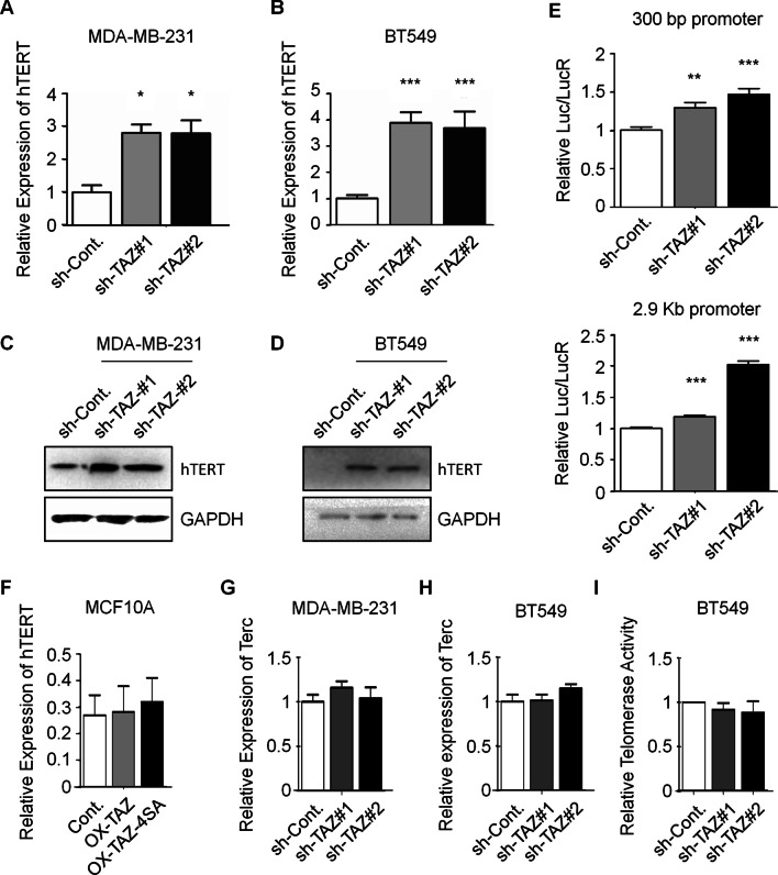
Results: By knocking down the expression of TAZ in TNBC cells, we found, for the first time, that TAZ is essential for the maintenance of telomeres in TNBC cells. Moreover, loss of TAZ causes senescence phenotype of TNBC cells. The observed extremely shortened telomeres in late passages of TAZ knocked down cells correlate with an elevated hTERT expression, reductions of shelterin proteins, and an activated DNA damage response pathway. Our data also showed that depletion of TAZ results in overexpression of TERRAs, which are a group of telomeric repeat-containing RNAs and regulate telomere length and integrity. Furthermore, we discovered that TAZ maintains telomere length of TNBC cells likely by facilitating the expression of Rad51C, a crucial element of homologous recombination pathway that promotes telomere replication.

Conclusions: This study supports the notion that TAZ is an oncogenic factor in TNBC, and further reveals a novel telomere-related pathway that is employed by TAZ to regulate TNBC.

Keywords: Rad51C; Shelterin proteins; TAZ; TERRAs; TNBC; Telomere.


Conflict of interest statement

The authors declare that they have no competing financial interests.

Figures

Fig. 1
Fig. 1
Knockdown of the expression of TAZ causes cell senescence in TNBC cells. A qPCR analysis in different breast cancer cell lines to indicate the mRNA levels of TAZ. B Western blot analysis in different breast cancer cell lines to indicate the protein levels of TAZ. Quantification of Western blots is shown in Additional file 1: Fig. S5A. C Western blot analysis in a control shRNA-transfected (sh-Cont.) and two separate TAZ-specific shRNAs-transfected (sh-TAZ) MDA-MB-231 cells to indicate the protein levels of TAZ and P21. Quantification of Western blots is shown in Additional file 1: Fig. S5B. D Cell viability assays were performed in the indicated MDA-MB-231 cells. E, Cell cycle assays were performed in the indicated MDA-MB-231 cells. F BrdU incorporation assays were performed in the indicated MDA-MB-231 cells. G Senescence-associated β-galactosidase activity staining analyses were performed in the indicated MDA-MB-231 cells. H qPCR analysis in the indicated MDA-MB-231 cells to indicated the mRNA levels of multiple SASP (senescence-associated secretory phenotype) marker genes. Data are presented as mean ± SEM. At least three repeats were carried out for each test. The p values of cell viability assays were determined by two-way ANOVA followed by Bonferroni post test. All other p values were determined by one-way ANOVA followed by Tukey’s multiple-comparisons. NS, not significant, *p < 0.05, **p < 0.01, ***p < 0.001
Fig. 2
Fig. 2
TAZ is essential for telomere maintenance in TNBC cells. A Western blot analysis in the indicated MDA-MB-231 cells to indicate the depletion efficiency of TAZ expression. Quantification of Western blots is shown in Additional file 1: Fig. S5C. B Telomere-specific qPCR analysis in the indicated MDA-MB-231 cells to determine the relative telomere length. C Southern analysis of TRFs in the indicated MDA-MB-231 cells to determine the relative telomere length. D Western blot analysis in the indicated BT549 cells to indicate the protein levels of TAZ. Quantification of Western blots is shown in Additional file 1: Fig. S5D. E Telomere-specific qPCR analysis in the indicated BT-549 cells to analyze the relative telomere length. F Southern analysis of TRFs in the indicated BT549 cells to determine the relative telomere length. G Western blot analysis in BT549 cells overexpressing wild type TAZ and its activated mutant (TAZ-4SA) with antibodies as indicated. Quantification of Western blots is shown in Additional file 1: Fig. S5E. H Telomere-specific qPCR analysis to determine the relative telomere length in the indicated BT549 cells. I Western blot analysis in T47D cells overexpressing wild type TAZ and its activated mutant (TAZ-4SA) with antibodies as indicated. J Telomere-specific qPCR analysis to determine the relative telomere length in the indicated T47D cells. K Western blot analysis in MCF10A cells overexpressing wild type TAZ and its activated mutant (TAZ-4SA) with antibodies as indicated. L Telomere-specific qPCR analysis to determine the relative telomere length in the indicated MCF10A cells. Data are presented as mean ± SEM. At least three repeats were carried out for each test. The p values were determined by one-way ANOVA followed by Tukey’s multiple-comparisons. NS, not significant, *p < 0.05, **p < 0.01, ***p < 0.001
Fig. 3
Fig. 3
Depletion of TAZ increases the expression of hTERT in TNBC cells. A and B qPCR analyses to indicate the mRNA levels of hTERT in indicated MDA-MB-231 and BT549 cells. C and D Western blot analyses in the indicated MDA-MB-231 and BT549 cells to examine the protein levels of hTERT. Quantification of Western blots is shown in Additional file 1: Fig. S5F. E Luciferase reporter assays were performed to test the activities of 300 bp and 2.9 kb hTERT promoter fragments in the indicated MDA-MB-231 cells. F qPCR analyses to indicate the mRNA levels of hTERT in indicated MCF10A cells. G and H qPCR analyses to indicate the mRNA levels of Terc in indicated MDA-MB-231 and BT549 cells. I TRAP analysis to measure telomerase activities in BT549 cells transfected as indicated. Data are presented as mean ± SEM. At least three repeats were carried out for each test. The p values were determined by one-way ANOVA followed by Tukey’s multiple-comparisons. NS, not significant, *p < 0.05, **p < 0.01, ***p < 0.001
Fig. 4
Fig. 4
Knockdown of TAZ results in the deprotection of telomeres and the activation of DDR. A Western blot analysis of shelterin proteins in TAZ-depleted MDA-MB-231 cells with long-term approaches (infected with shRNA and cultured > 6 passages). Quantification of Western blots is shown in Additional file 1: Fig. S5G. B Western blot analysis of shelterin proteins in long-term TAZ knockdown (infected with shRNA and cultured > 6 passages) BT549 cells. Quantification of Western blots is shown in Additional file 1: Fig. S5H. C Immunofluorescence assay of TRF2 in BT549 and MDA-MB-231 cells treated as indicated. D Western blot analysis of the DDR pathway proteins in MDA-MB-231 and BT549 cell with long-term TAZ knockdown. Quantification of Western blots is shown in Additional file 1: Fig. S5I. E Immunofluorescence assay of p-BRCA1 in BT-549 cells with long-term TAZ knockdown. F IF-FISH assays stained with 53BP1 antibody and Cy3-conjugated telomere PNA probes were performed in the indicated BT549 cells. Co-localizing events indicate telomere dysfunction induced foci (TIFs). The percentage of cells with more than 3 TIFs were analyzed. 100 cells were counted for each experiment. G Immunofluorescence assays of TRF2 and γ-H2AX in the indicated BT549 cells. Co-localizing events indicate telomere dysfunction induced foci (TIFs). The percentage of cells with more than 3 TIFs were analyzed. 100 cells were counted for each experiment
Fig. 5
Fig. 5
Knockdown of TAZ inhibits Rad51C transcription, and TAZ maintains telomere length through Rad51C. A Western blot analysis of TAZ and Rad51C protein levels in MDA-MB-231 and BT549 cells as indicated. Quantification of Western blots is shown in Additional file 1: Fig. S5J. B qPCR analyses to determine the mRNA levels of Rad51C in MDA-MB-231 and BT549 cells as indicated. C Western blot analysis in BT549 cells treated as indicated to examine the protein levels of Rad51C. Quantification of Western blots is shown in Additional file 1: Fig. S5K. D Telomere-specific qPCR analysis to determine the relative telomere length in the indicated BT549 cells. E Immunofluorescence assays of TRF2 and γ-H2AX in the indicated BT549 cells. Co-localizing events indicate telomere dysfunction induced foci (TIFs). The percentage of cells with more than 3 TIFs were analyzed. 100 cells were counted for each experiment. F Western blot analyses in BT549 cells as indicated. Quantification of Western blots is shown in Additional file 1: Fig. S5L. G Telomere-specific qPCR analysis to determine the relative telomere length in the indicated BT549 cells. H Western blot analyses in BT549 cells and MDA-MB-231 treated as indicated to examine the protein levels of TAZ and Rad51C. Quantification of Western blots is shown in Additional file 1: Fig. S5M. I Telomere-specific qPCR analysis to determine the relative telomere length in the indicated BT549 and MDA-MB-231 cells. J Southern analysis of TRFs in the indicated BT549 and MDA-MB-231 cells to determine the relative telomere length. Data are presented as mean ± SEM. At least three repeats were carried out for each test. Statistical analyses were performed with unpaired Student's t-test between two groups and one-way ANOVA followed by Tukey’s multiple-comparisons for multiple groups. NS, not significant, *p < 0.05, **p < 0.01, ***p < 0.001
Fig. 6
Fig. 6
Rad51C is a direct YAP/TAZ-TEAD target gene. A TEAD4 binding signals in the promoter of Rad51C from ENCODE ChIP-seq database (https://www.encodeproject.org) were analyzed from A549 (lung cancer), H1 (human embryonic stem cells), HCT-116 (colon cancer), MCF-7 (breast cancer), and SK-N-SH (neuroblastoma) cells. B Western blot analysis of TAZ and TEAD4 protein levels in BT549 cells as indicated. Quantification of Western blots is shown in Additional file 1: Fig. S5N. C ChIP analyses in the indicated BT549 cells were performed to determine the enrichment of TRAD4 on the Rad51C promoter region. IgG was used as a negative control. D Luciferase reporter assays were performed to test the activities of Rad51C promoter fragments in the indicated BT549. Data are presented as mean ± SEM. At least three repeats were carried out for each test. The p values were determined by Student's t-test. NS, not significant, *p < 0.05, **p < 0.01, ***p < 0.001
Fig. 7
Fig. 7
Working model of TAZ regulating telomere length in TNBC cells. In TAZ-knocked down TNBC cells, the binding of TAZ-TEADs to the promoter of Rad51C was decreased, and the transcription of Rad51C was further impaired. Lack of Rad51C causes the telomere shortening and impaired telomere functions. The protein levels of shelterin components is decreased, along with increased telomere-induced DNA damage foci (TIF) and DDR activation. Moreover, the increased levels of TERRAs in TAZ-deficient cells may also contribute to the observed telomere loss phenotype

References

    1. Yin L, et al. Triple-negative breast cancer molecular subtyping and treatment progress. Breast Cancer Res. 2020;22(1):61. doi: 10.1186/s13058-020-01296-5. - DOI - PMC - PubMed
    1. Zanconato F, Cordenonsi M, Piccolo S. YAP/TAZ at the roots of cancer. Cancer Cell. 2016;29(6):783–803. doi: 10.1016/j.ccell.2016.05.005. - DOI - PMC - PubMed
    1. Zhou X, Lei QY. Regulation of TAZ in cancer. Protein Cell. 2016;7(8):548–561. doi: 10.1007/s13238-016-0288-z. - DOI - PMC - PubMed
    1. Chan SW, et al. A role for TAZ in migration, invasion, and tumorigenesis of breast cancer cells. Can Res. 2008;68(8):2592–2598. doi: 10.1158/0008-5472.CAN-07-2696. - DOI - PubMed
    1. Cordenonsi M, et al. The Hippo transducer TAZ confers cancer stem cell-related traits on breast cancer cells. Cell. 2011;147(4):759–772. doi: 10.1016/j.cell.2011.09.048. - DOI - PubMed

Publication types

MeSH terms