. 2021 Sep 7;7(1):81.
doi: 10.1038/s41421-021-00316-8.

Generation of human blastocyst-like structures from pluripotent stem cells

Affiliations

Generation of human blastocyst-like structures from pluripotent stem cells

Yong Fan et al. Cell Discov. .

Abstract

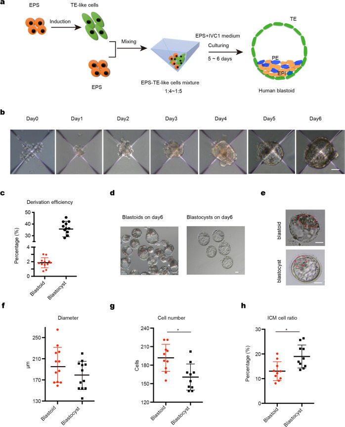
Human blastocysts are comprised of the first three cell lineages of the embryo: trophectoderm, epiblast and primitive endoderm, all of which are essential for early development and organ formation. However, due to ethical concerns and restricted access to human blastocysts, a comprehensive understanding of early human embryogenesis is still lacking. To bridge this knowledge gap, a reliable model system that recapitulates early stages of human embryogenesis is needed. Here we developed a three-dimensional (3D), two-step induction protocol for generating blastocyst-like structures (EPS-blastoids) from human extended pluripotent stem (EPS) cells. Morphological and single-cell transcriptomic analyses revealed that EPS-blastoids contain key cell lineages and are transcriptionally similar to human blastocysts. Furthermore, EPS-blastoids are similar with human embryos that were cultured for 8 or 10 days in vitro, in terms of embryonic structures, cell lineages and transcriptomic profiles. In conclusion, we developed a scalable system to mimic human blastocyst development, which can potentially facilitate the study of early implantation failure that induced by developmental defects at early stage.


Conflict of interest statement

The authors declare no competing interests.

Figures

Fig. 1
Fig. 1. Induction of human blastoids under 3D two-step condition.
a Schematic of human blastoid formation. EPS cells were firstly induced to TE-like cells, and then TE-like cells were mixed with EPS cells and seeded together to AgreeWell on day 0. The aggregates further differentiated and organized into a human EPS-blastoid. b Phase-contrast images of human aggregates in the indicated days showing the formation of human blastoids from day 0 to day 6. Scale bar = 5 μm. c Derivation efficiency of human blastoids is about 1.9% that significantly lower than the developmental efficiency of human blastocysts. d Phase-contrast image of human blastoids on day 6, Scale bar = 50 μm. e Phase-contrast image of human EPS-blastoid (upper) and human blastocyst (lower). Red line indicated inner cell mass (ICM) of the structure and the outer layer cells represented trophoblast cells (TE). Scale bar = 50 μm. fh Mean diameter (f), total cell number (g), and ICM cell ratio (h) were quantified between human EPS-blastoids and blastocyst. n = 30 EPS-blastoids, n = 30 blastocyst. Data in c, data are means ± SD (n = 12 blastoids). **P < 0.001. Data in e, data are means ± SD (n = 12 blastoids). P > 0.05. Data in f and g, data are means ± SD (n = 12 blastoids). *P < 0.05. Data in h, data are numbers (n = 40 blastoids, 40 blastocysts), **P < 0.001.
Fig. 2
Fig. 2. Expression of specific development-related markers.
af Immunofluorescence staining of human EPS-blastoids for EPI lineage marker (OCT4), TE lineage markers (CK8 and GATA2/3), and PE lineage marker (GATA6), Scale bar = 50 μm. g Model of immunofluorescence staining of human blastocyst for OCT4 in EPI, GATA6 in PE, GATA2/3, and CK8 in TE lineages, Scale bar = 50 μm. hj Quantification of the percentage of human blastoids with correct allocation of OCT4 and CK8 (h), GATA6 (i), and all three markers (j). Data in i, data are means ± SD (n = 172 blastoids). *P < 0.05. Data in i, j, data are means ± SD (n = 131 blastoids).
Fig. 3
Fig. 3. Landscape of transcriptome in human blastoids on day 6.
a A UMAP plot 10,933 cells from human blastoids showing the cells in blastoids were divided into 4 major clusters on day 6. EPI/ICM, PE, TE, and IM (intermediate) subgroups were determined according to the lineage-specific markers. The cells in IM subgroups express all three lineages markers or some uncertain genes. b Heat map of lineage signature genes expression on day 6. c UMAP projection of integrated datasets showing EPI-, PE-, and TE-like cells (and IM clusters) together with EPI, PE, and TE cells of blastocysts from a previous published study. d Dot plot indicating the markers of EPI, PE, and TE lineage markers. e Overlapping genes expression between a previous study and this study was performed by heat map and GO term analysis.
Fig. 4
Fig. 4. Landscape of transcriptome in human embryo-like structures from blastoids on day 8 and day 10.
ad Immunofluorescence staining of EPI marker OCT4 and PE marker GATA6 in Day 8 (a) and Day 10 (b) embryonic structures. A UMAP plot analysis revealed 5 clusters in embryonic structures on day 8 (c) and day 10 (d), identified with assigned cluster names. e UMAP projection of integrated datasets showing our results of embryonic structures on day 8, day 10, and previous studies, (data of 7–14 d.p.f.). f Heatmap of lineage-specific genes overlapped between a previous study (data of 7–14 d.p.f. embryos) and this study on days 8 and 10.

References

    1. Shahbazi MN. Mechanisms of human embryo development: from cell fate to tissue shape and back. Development. 2020;147:dev190629. doi: 10.1242/dev.190629. - DOI - PMC - PubMed
    1. Rossant J, Tam PPL. New insights into early human development: lessons for stem cell derivation and differentiation. Cell Stem Cell. 2017;20:18–28. doi: 10.1016/j.stem.2016.12.004. - DOI - PubMed
    1. Wamaitha SE, Niakan KK. Human pre-gastrulation development. Curr. Top. Dev. Biol. 2018;128:295–338. doi: 10.1016/bs.ctdb.2017.11.004. - DOI - PubMed
    1. Niakan KK, Han J, Pedersen RA, Simon C, Pera RA. Human pre-implantation embryo development. Devevelopment. 2012;139:829–841. - PMC - PubMed
    1. Nakamura T, et al. A developmental coordinate of pluripotency among mice, monkeys and humans. Nature. 2016;537:57–62. doi: 10.1038/nature19096. - DOI - PubMed