. 2021 Sep 6;11(1):17705.
doi: 10.1038/s41598-021-97162-1.

HSPA9/Mortalin mediates axo-protection and modulates mitochondrial dynamics in neurons

Affiliations

HSPA9/Mortalin mediates axo-protection and modulates mitochondrial dynamics in neurons

Cécile A Ferré et al. Sci Rep. .

Abstract

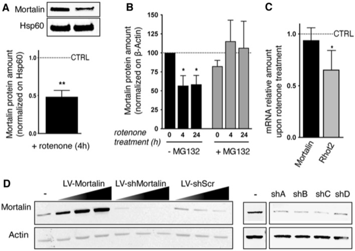
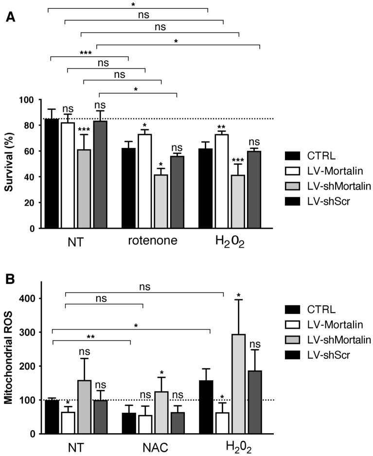
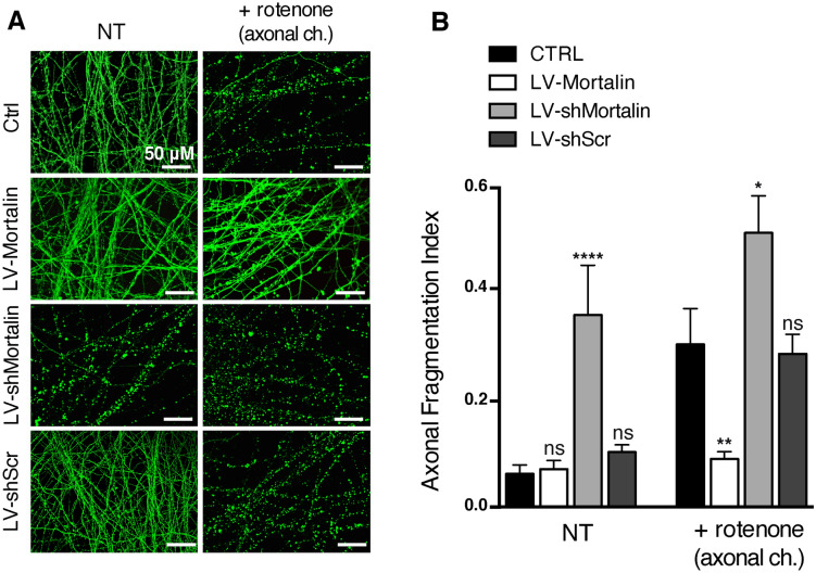
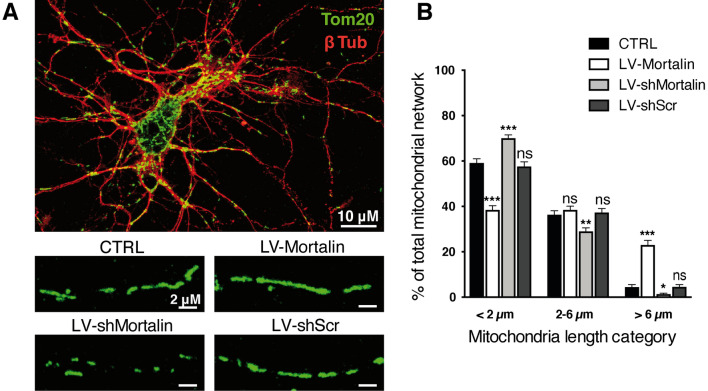
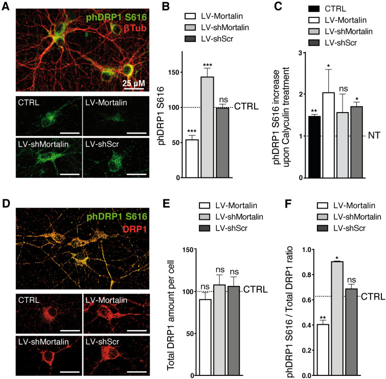
Mortalin is a mitochondrial chaperone protein involved in quality control of proteins imported into the mitochondrial matrix, which was recently described as a sensor of neuronal stress. Mortalin is down-regulated in neurons of patients with neurodegenerative diseases and levels of Mortalin expression are correlated with neuronal fate in animal models of Alzheimer's disease or cerebral ischemia. To date, however, the links between Mortalin levels, its impact on mitochondrial function and morphology and, ultimately, the initiation of neurodegeneration, are still unclear. In the present study, we used lentiviral vectors to over- or under-express Mortalin in primary neuronal cultures. We first analyzed the early events of neurodegeneration in the axonal compartment, using oriented neuronal cultures grown in microfluidic-based devices. We observed that Mortalin down-regulation induced mitochondrial fragmentation and axonal damage, whereas its over-expression conferred protection against axonal degeneration mediated by rotenone exposure. We next demonstrated that Mortalin levels modulated mitochondrial morphology by acting on DRP1 phosphorylation, thereby further illustrating the crucial implication of mitochondrial dynamics on neuronal fate in degenerative diseases.


Conflict of interest statement

The authors declare no competing interests.

Figures

Figure 1
Figure 1
Modulation of Mortalin levels in primary cultured neurons. (A) Mortalin is down-regulated in neurons upon treatment with rotenone. Rat embryonic primary cortical neurons (DIV 12) were treated or not with 10 nM rotenone (+ rotenone) for 4 h and protein extracts were prepared from mitochondria-enriched fractions. Mortalin amounts were assessed by Western blot, using the mitochondrial chaperone Hsp60 or ß-Actin as loading controls. The graph represents the normalized ratio relative to untreated neurons and is expressed as the mean ± SD of 4 independent experiments. (B) Mortalin loss upon rotenone treatment is due to proteasomal degradation. Neurons (DIV 12) were treated or not with 10 nM rotenone for 4 or 24 h with or without MG132. Neuronal extracts were then analyzed by Western blotting for Mortalin protein quantification. ß-Actin was used as a loading control. The graph represents means ± SD of Mortalin amounts in treated neurons, normalized on control, untreated ones (100%), in 3 independent experiments. (C) Mortalin RNA levels are not affected by rotenone treatment. RNA was extracted from neurons treated or not with 10 nM rotenone for 4 h. qRT-PCR experiments were performed to determine mRNA levels for Mortalin and Rhot2 (Miro2 gene), as well as for Mdh1, a housekeeping gene. The ratios of Mortalin and Rhot2 mRNA levels (normalized on Mdh1) in rotenone treated vs. untreated neurons were calculated and data are presented as means ± SD of 4 independent experiments. (D) Modulation of Mortalin expression by transduction with lentiviral vectors (LV). Overexpression was achieved with a LV bearing an expression cassette for the rat Mortalin gene. Mortalin down-regulation was driven by cell transduction with lentiviral vectors (numbered sh A to D) expressing a set of specific 29-mer shRNA directed against Mortalin (shMortalin), or a control scramble sequence (shScr, Origene TR704140). Different multiplicities of transduction (1, 2, 4) were used to test the percentage of over- or under-expression. WB are presented as cropped parts of full blots displayed as supplemental information. DIV: days in vitro. *: p < 0.05, **p < 0.01, by Mann–Whitney non parametric t-test.
Figure 2
Figure 2
Mortalin levels control neuronal survival against stress induced by rotenone and H2O2 and mitochondrial accumulation of ROS. Rat embryonic primary cortical neurons were transduced on DIV 3 with lentiviral vectors (LV) to induce over- (+ 100%, LV-Mortalin) or under- (− 50%, LV-shMortalin, clone A) expression of Mortalin. A lentiviral vector expressing a shRNA with a scramble sequence was used as a control (LV-shScr). (A) On DIV 11, neurons were treated or not with 10 nM rotenone for 24 h, or with hydrogen peroxide (H2O2, 100 μM) and then processed the day after. Percentages of cells showing pyknosis (revealed by DAPI staining) are presented as means ± SD of 4 independent experiments. (B) On DIV 11, neurons were treated or not with 100 μM NAC or H2O2. After 24 h, mitochondrial ROS accumulation was measured using the MitoSOX dye. Values were normalized using fluorometric values obtained for untreated and non-transduced neurons for each experiment. Results are presented as means ± SD of 3 independent experiments. ****p < 10−4, by 1-way ANOVA with Sidak’s multiple comparison post-hoc test.
Figure 3
Figure 3
Mortalin expression controls axonal fate against a distal oxidative stress insult. Rat embryonic primary cortical neurons were grown in microfluidic devices for 12 DIV. Neurons were transduced on DIV 3 with lentiviral vectors (LV) to induce over- (LV-Mortalin) or under- (LV-shMortalin, clone A) expression of Mortalin. A lentiviral vector expressing a shRNA with scramble nucleotide sequence was used as a control (LV-shScr). On DIV 11, neurons were treated or not with 1 μM rotenone applied in the axonal chamber (+ rotenone) for 24 h and fixed on DIV 12. (A) Pictures of βIII-tubulin staining were randomly taken within axonal chambers (4 by condition) and the axonal fragmentation indexes were measured for each picture as the number of axonal dots per unit of tubulin staining. (B) Results are presented as means ± SD of 4 to 10 independent microfluidic devices, from 4 independent neuronal cultures. *p < 0.05, **p < 0.01, ****p < 10−4, by 1-way ANOVA with Sidak’s multiple comparison post-hoc test.
Figure 4
Figure 4
Mortalin modulates mitochondrial morphology in neuronal extensions. Rat embryonic primary cortical neurons were transduced on DIV 3 with lentiviral vectors (LV) to induce over- (LV-Mortalin) or under- (-LV-shMortalin, clone A) expression of Mortalin. A lentiviral vector expressing a shRNA with a scramble sequence was used as a control (LV-shScr). (A) Neurons were fixed on DIV 12 and both neuronal and mitochondrial networks were revealed by staining with, respectively, Tom20 and βIII-tubulin (βIII-Tub). Pictures were randomly taken and mitochondrial lengths were measured in all neuronal extensions. (B) For each neuron, the sum of the lengths of all measured mitochondria was calculated (mitochondrial network), within which the relative proportions of short (< 2 m), medium (2–6 m) and long (> 6 m) mitochondria were considered. The graph represents means ± SD of 18 to 40 neuronal mitochondrial networks, from 4 independent neuronal preparations. *p < 0.05, **p < 0.01, ***p < 0.001, by 2-way ANOVA, Kruskal Wallis test and Dunn’s multiple comparisons as post-hoc tests.
Figure 5
Figure 5
Mortalin impacts mitochondrial morphology by modulating Drp1-S616 phosphorylation. Rat embryonic primary cortical neurons were transduced on DIV 3 with lentiviral vectors (LV) to induce over- (LV-Mortalin) or under- (LV-shMortalin, clone A) expression of Mortalin. A lentiviral vector expressing a shRNA with a scramble sequence was used as a control (LV-shScr). Neurons were then fixed on DIV 12 and the active form of Drp1, phosphorylated on serine 616, was revealed by pDrp1-S616 staining. (AC) The neuronal network was visualized with the neuronal marker βIII-tubulin (βIII-Tub). On each randomly taken picture (A), the total amount of pDrp1 was measured (integrated staining density, Image J software) and normalized on the βIII-tubulin staining area. For each experiment, 4 pictures were taken per condition and the corresponding mean ratios were normalized relative to control, non-transduced neurons. (B). The same was done upon treating neurons with Calyculin-A for 30 min before fixation, in order to inhibit phosphatase activity (C). The results are presented as means ± SD of 4 experiments. (D–F) Neurons were co-strained with antibodies directed against total Drp1 and pDrp1-S616 (D). For each randomly taken picture, we evaluated the total and phosphorylated protein amounts in βIII-Tub + cells, using the integrated staining density (Image J software). The values obtained for total Drp1 were normalized on the signal for non-transduced neurons in each experiment and are presented as means ± SD of 3 independent experiments (E). Ratios of the integrated staining densities for pDrp1-S616 on total Drp1 were also measured and normalized on the ratio obtained for un-transduced cells and presented as means ± SD of 3 independent experiments (F). **p < 0.01, by Mann–Whitney non parametric test.

References

    1. Vickers JC, et al. Axonopathy and cytoskeletal disruption in degenerative diseases of the central nervous system. Brain Res. Bull. 2009;80:217–223. doi: 10.1016/j.brainresbull.2009.08.004. - DOI - PubMed
    1. Raff MC, Whitmore AV, Finn JT. Axonal self-destruction and neurodegeneration. Science. 2002;296:868–871. doi: 10.1126/science.1068613. - DOI - PubMed
    1. Adalbert R, Coleman MP. Review: Axon pathology in age-related neurodegenerative disorders. Neuropathol. Appl. Neurobiol. 2013;39:90–108. doi: 10.1111/j.1365-2990.2012.01308.x. - DOI - PubMed
    1. Coleman MP, Freeman MR. Wallerian degeneration, wld(s), and nmnat. Annu. Rev. Neurosci. 2010;33:245–267. doi: 10.1146/annurev-neuro-060909-153248. - DOI - PMC - PubMed
    1. Babetto E, et al. Targeting NMNAT1 to axons and synapses transforms its neuroprotective potency in vivo. J. Neurosci. 2010;30:13291–13304. doi: 10.1523/JNEUROSCI.1189-10.2010. - DOI - PMC - PubMed

Publication types