Review
. 2021 Aug 19:15:721186.
doi: 10.3389/fncir.2021.721186. eCollection 2021.

Corticothalamic Pathways in Auditory Processing: Recent Advances and Insights From Other Sensory Systems

Affiliations
Review

Corticothalamic Pathways in Auditory Processing: Recent Advances and Insights From Other Sensory Systems

Flora M Antunes et al. Front Neural Circuits. .

Abstract

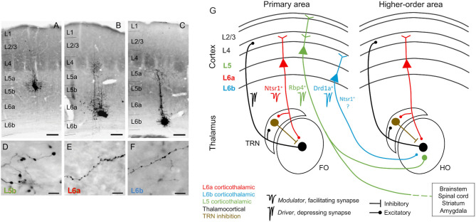
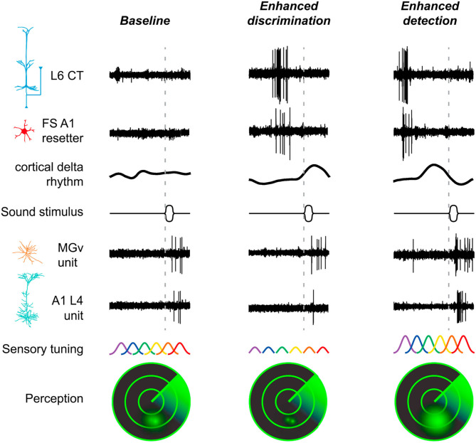
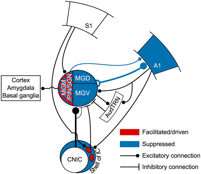
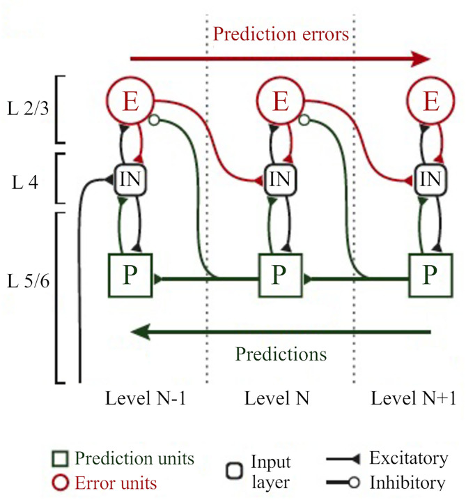
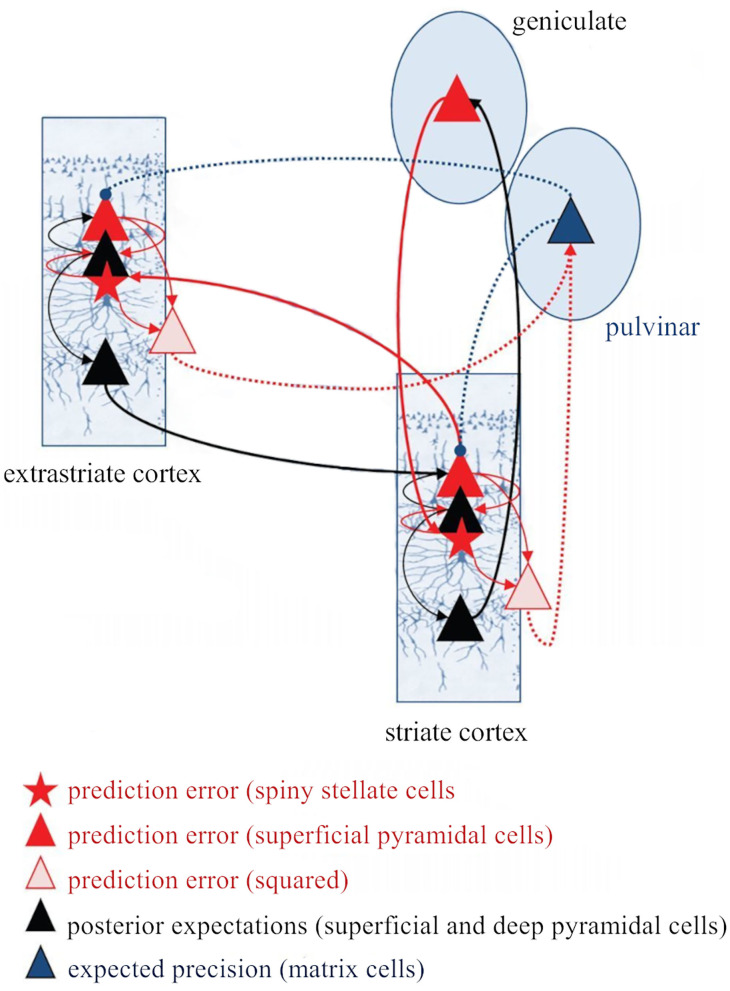
The corticothalamic (CT) pathways emanate from either Layer 5 (L5) or 6 (L6) of the neocortex and largely outnumber the ascending, thalamocortical pathways. The CT pathways provide the anatomical foundations for an intricate, bidirectional communication between thalamus and cortex. They act as dynamic circuits of information transfer with the ability to modulate or even drive the response properties of target neurons at each synaptic node of the circuit. L6 CT feedback pathways enable the cortex to shape the nature of its driving inputs, by directly modulating the sensory message arriving at the thalamus. L5 CT pathways can drive the postsynaptic neurons and initiate a transthalamic corticocortical circuit by which cortical areas communicate with each other. For this reason, L5 CT pathways place the thalamus at the heart of information transfer through the cortical hierarchy. Recent evidence goes even further to suggest that the thalamus via CT pathways regulates functional connectivity within and across cortical regions, and might be engaged in cognition, behavior, and perceptual inference. As descending pathways that enable reciprocal and context-dependent communication between thalamus and cortex, we venture that CT projections are particularly interesting in the context of hierarchical perceptual inference formulations such as those contemplated in predictive processing schemes, which so far heavily rely on cortical implementations. We discuss recent proposals suggesting that the thalamus, and particularly higher order thalamus via transthalamic pathways, could coordinate and contextualize hierarchical inference in cortical hierarchies. We will explore these ideas with a focus on the auditory system.

Keywords: corticothalamic circuits; feedback loops; hierarchical inference; reticular thalamic nucleus; transthalamic pathways.


Conflict of interest statement

The authors declare that the research was conducted in the absence of any commercial or financial relationships that could be construed as a potential conflict of interest.

Figures

Figure 1
Figure 1
Corticothalamic neurons and circuits. (A–C) Photomicrographs showing BDA microdeposits in L5b, L6a, and L6b neurons of primary somatosensory barrel field (D–F), and their respective axonal arborizations in (higher order) posterior thalamic nucleus. Cortical pyramidal neurons differ in the morphology of their axonal varicosities. Scale bars: A–C = 150 μm, D–F = 10 μm. (G) Schematic and simplified view of corticothalamic and thalamocortical circuits, based on information from several sensory systems (auditory, visual, and somatosensory). Red, L6a CT projections, modulator (Ntsr1+): feedback projections that send collaterals to the TRN, a GABAergic nucleus that provides inhibition to the thalamus (brown). Black, thalamocortical, feedforward projections that form reciprocal loops with L6a feedback projections. Green, L5 CT projections, driver (Rbp4+): project non-reciprocally to a hierarchically higher order thalamic nucleus and form part of transthalamic corticocortical pathways; they are collaterals from long-range axons that project to other subcortical centers (e.g., brainstem, spinal cord, striatum, and amygdala). Blue, L6b CT projections, driver (Drd1a+): project non-reciprocally to a hierarchically higher order thalamic nucleus; it is unknown if Drd1a+ and Ntsr1+ neurons in L6b are overlapping or distinct neuronal populations. As insets, examples of specific markers from Cre mouse lines that can be used to selectively target neurons of each CT circuit [Ntsr1-Cre, Drd1a-Cre, and Rbp4-Cre mice, for L6a (red), L6b (blue), and L5 (green) CT neurons]. FO, first order thalamic nuclei; HO, higher order thalamic nuclei; TRN, thalamic reticular nucleus; L2/3, cortical layers 2 and 3; L4, cortical layer 4; L5, cortical layer 5; L6a, cortical layer 6a; L6b, cortical layer 6b; Photomicrographs (A–F) after Hoerder-Suabedissen et al. (2018).
Figure 2
Figure 2
The cortex exerts suppressive and facilitating influences in neurons of the auditory thalamus. Examples of single-unit responses to auditory stimulation in the MGB of the anesthetized rat before, during, and after AC deactivation by cooling. (A) A neuron localized to the MGM that receives suppressive influences from the AC. The frequency response areas (first row), and the responses of the neuron to the oddball paradigm (second-fourth rows), in the control, cool and recovery conditions. The oddball paradigm was used to elicit SSA in these neurons. Briefly, the oddball paradigm consisted of a sequence of a repetitive stimulus (standard; 90% probability) that was infrequently interrupted by a different stimulus (deviant; 10% probability. The standard (blue) and the deviant (red) stimulus were pure tones selected from within the frequency response area of the neuron. Two blocks of 400 trials each (middle panels) were presented in which the standard and deviant frequencies were reversed (second panel, first block: f1/f2 as standard/deviant; third panel, second block: f2/f1 as standard/deviant). Dot rasters show individual spikes to the deviant and standard (red and blue dots, respectively), in the three conditions for the two stimulus presentation blocks (stacked along the y-axis; repetition rate 4 Hz; stimulus duration: 75 ms, black horizontal lines under the plots). PSTHs (last row) show the number of spikes/stimulus (bin duration: 3 ms) averaged over the two blocks [(f1+f2)/2; blue line is standard, red line is deviant]. The CSI calculated for each condition is noted as an inset on the PSTHs. The CSI quantifies the amount of SSA and is calculated as CSI = [d(f1)+d(f2)-s(f1)-s(f2)]/[d(f1)+d(f2)+s(f1)+s(f2)], where d(fi) and s(fi) are responses (# spikes/stimulus) to frequency fi when deviant or standard, respectively (0 ≤ CSI ≤ 1). Higher CSI values, higher SSA. (B) Responses of another neuron localized to the MGM that receives facilitatory influences from the AC, presented as in (A). AC, auditory cortex; MGB, medial geniculate body; MGM, medial subdivision of the medial geniculate body; SSA, stimulus specific adaptation; CSI, common SSA index; f1, frequency 1; f2, frequency 2. Adapted from Antunes and Malmierca (2011).
Figure 3
Figure 3
Non-SSA neurons primarily receive facilitatory influences from the AC. Example of a neuron recorded from the MGV that was facilitated by the AC, presented as in Figure 2. The neuron responds consistently to both the standard and the deviant over the trials, i.e., it does not show SSA as confirmed by the low CSI value (~0). The CSI value was not significantly changed by AC deactivation. AC, auditory cortex; MGV, ventral subdivision of the medial geniculate body; CSI, common SSA index. Adapted from Antunes and Malmierca (2011).
Figure 4
Figure 4
The gain exerted by the cortex in auditory thalamic neurons depends on their ability to signal a deviance from previous stimulation context. Scatterplots of the CSI (control condition) vs. the difference in firing rate between the control and cool conditions (spikes/stimulus difference) in response to the standard (upper panel) and deviant stimulus (lower panel), for each neuron. Blue, green, and red dots represent the neurons that were localized to the ventral (n = 12), dorsal (n = 24), and medial (n = 9) subdivisions of the MGB, respectively (n = 45, neurons that were localized to one of the three MGB subdivision). Gray dots represent MGB neurons that were not localized to a specific MGB subdivisions (n = 3). In both plots, positive values (above the horizontal line at the origin) indicate a reduction in firing rate with cortical deactivation (neurons receive facilitatory cortical influences), whereas negative values (bellow the horizontal line) indicate an increment in firing rate with cortical deactivation (neurons receive suppressive influences from the cortex). The difference in firing rate was inversely correlated with CSI for both standard and deviant stimuli. The slopes of the standard and deviant regression lines are not significantly different from each other [ANCOVA: main effect of stimuli, F(1,92) = 1.89, p = 0.172; main effect of CSI, F(1,92) = 43.27, p = 0; interaction, F(1,92) = 0.23, p = 0.634; n = 48], indicating that the correlation coefficients between standard and deviant are not different. The AC differentially affects the discharge rate of neurons depending on their SSA level. Neurons without SSA are mainly facilitated, whereas some neurons with high SSA are suppressed by the cortex. AC, auditory cortex; MGB, medial geniculate body; MGV, ventral subdivision of the MGB; MGD, dorsal subdivision of the MGB; MGM, medial subdivision of the MGB; SSA, stimulus specific adaptation; CSI, common SSA index; Adapted from Antunes and Malmierca (2011).
Figure 5
Figure 5
L6 CT neurons are involved in the behavioral switch between sound detection and discrimination. This scheme summarizes the findings by Guo et al. (2017) who demonstrate the participation of L6 CT neurons (Ntsr1+), via both their intracortical and corticothalamic connections to perceptual modes of enhanced detection or discrimination. Left column, in a baseline condition with low activity in L6 CT neurons and fast-spiking interneurons (FS resetters), the sound-evoked activity in MGV and L4 cortical neurons is moderate. FS resetters are activated following intense firing of L6 CT neurons. Activity of FS resetters increases the power and resets the phase of low frequency rhythms. Middle column, at a short delay period following intense activity of L6 CT and FS resetter neurons, the delta-theta rhythm is at a positive, low excitability phase, and sound-evoked activity is suppressed in A1 but not in the MGV, which favors tone discrimination at the expense of sound sensitivity (the reduced excitability of cortical neurons sharpens frequency tuning). Right column, at longer delays following L6 CT activity, the phase of the cortical rhythm has rotated to a negative, high excitability phase, and sound-evoked activity is enhanced both in A1 and MGV. The enhanced excitability of cortical neurons is expected to increase the overlap in sensory tuning between neighboring tuning regions, which favors tone detection at the expense of reduced tone discrimination. A1, primary auditory cortex; CT, corticothalamic; L6, cortical layer 6; L4, cortical layer 4; MGV, ventral subdivision of the medial geniculate body; FS, fast-spiking interneurons. Reproduced, with permission, from Guo et al. (2017).
Figure 6
Figure 6
L6b neurons project to higher order and avoid first order thalamic nuclei. Drd1a-Cre expression in fibers arising from neurons in L6b of the entire cortical mantle in adult mice (P35) visualized by tdTomato labeling (Drd1a-Cre::tdTom+ fibers). Projections from L6b avoid first order auditory and non-auditory thalamic nuclei such as (A) the anteroventral nucleus, (B) the ventral-posterior medial nucleus, (C) the lateral geniculate nucleus anteriorly, and (D) the MGV in the auditory thalamus. In contrast, they innervate heavily higher order thalamic nuclei such as the lateral dorsal nucleus, the posterior nucleus, the ventral medial nucleus, and the ventral anterior lateral complex. Fibers pass through TRN without apparent branching. Scale bar Scale bar = 500 μm. AD, anterodorsal nucleus; AV, anteroventral nucleus; CM, central medial nucleus; IMD, intermediodorsal nucleus; LD, lateral dorsal nucleus; LH, lateral habenula; LGd, dorsal lateral geniculate nucleus; LGv, ventral lateral geniculate nucleus; LP, lateral posterior nucleus; MD, mediodorsal nucleus; MG, medial geniculate nucleus; MH, medial habenula; Po, posterior nucleus; POL, posterior limiting nucleus; PVT, paraventricular nucleus; RE, nucleus reuniens; RH, rhomboid nucleus; SPF, subparafascicular nucleus; TRN, thalamic reticular nucleus; VAL, ventral anterior lateral complex; VM, ventral medial nucleus; VPL, ventral-posterior lateral nucleus; VPM, ventral-posterior medial nucleus. Reproduced from Hoerder-Suabedissen et al. (2018).
Figure 7
Figure 7
Auditory thalamus and midbrain bridging primary somatosensory cortex (S1) to primary auditory cortex (A1). This scheme summarizes the findings of Lohse et al. (2021) study. This study elegantly undisclosed the multisensory circuits by which the primary somatosensory cortex controls activity in the thalamocortical system in mice. In blue, regions of the auditory system (midbrain, thalamus and A1) that were suppressed to auditory stimulation (tones) by concurrent whisker stimulation (whisker deflection). Suppression occurred via a descending connection from S1 to neurons in the lateral shell of the inferior colliculus that project to the MGB. This resulted in a suppression of thalamocortical neurons and suppression of auditory activity in A1. Altogether, this forms a corticocolliculo-thalamocortical multisensory circuit by which somatosensory information exerts a dominance over auditory processing in A1. In red, some neurons in the medial sector of the auditory thalamus, including the MGM, had their auditory responses enhanced or driven with whisker stimulation. A direct connection arising in L5 of S1 to the medial sector of the MGB could mediate this enhancement. Because the MGM projects to cortical areas, including A1, this could form a transthalamic circuit bridging S1 to AC. However, this hypothesis needs further confirmation. A1, primary auditory cortex; AudTRN, auditory sector of the thalamic reticular nucleus; CNIC, central nucleus of the inferior colliculus; MGM/PIN/SGN, medial subdivision of the MGB/posterior intralaminar nucleus/suprageniculate nucleus; MGD, dorsal subdivision of the MGB; MGV, ventral subdivision of the MGB; S1, primary somatosensory cortex. Adapted from Lohse et al. (2021).
Figure 8
Figure 8
The classical implementation of predictive coding relies on the cortical hierarchy. A simplified hierarchical model based on the classical implementation of predictive coding (Bastos et al., 2012). Vertical dashed lines delimit hierarchically arranged cortical columns (left to right: bottom up). Prediction errors climb up the cortical hierarchy through the feedforward, bottom-up connections, whereas predictions are sent backwards (top-down) to suppress prediction error units of the levels below, via inhibitory connections. Superficial layers (L2/3) above the subcortical input layer (L4) carry prediction errors, whereas deep cortical layers (L5/6) carry predictions. Adapted, with permission, from Heilbron and Chait (2018) and Carbajal and Malmierca (2020).
Figure 9
Figure 9
The thalamus and the corticothalamic pathways as key players in the implementation of predictive coding. This scheme, proposed by Kanai et al. (2015) expands the classical implementation of predictive coding [Bastos et al. (2012), Shipp (2016)] as Figure 7 shows, because it incorporates the thalamus and the corticothalamic pathways (both L6a and L5 CT projections) as key players in its implementation. A very interesting aspect of this model is the inclusion of deep pyramidal cells (presumably L5 CT cells) that convey the squared prediction error (second-order forward connections) to enable the pulvinar (matrix cells) to estimate precision. These cells in the pulvinar send back projections to modulate the gain of superficial pyramidal cells in the cortex. Red, forward connections; Black, backward connections; Full lines, first order streams; Dashed lines, second order (precision-related) streams. CT, corticothalamic; L5, cortical layer 5; L6a, cortical layer 6a. Adapted from Kanai et al. (2015).

References

    1. Abbott L. F., Regehr W. G. (2004). Synaptic computation. Nature 431, 796–803. 10.1038/nature03010 - DOI - PubMed
    1. Adams R. A., Shipp S., Friston K. J. (2013). Predictions not commands: active inference in the motor system. Brain Struct. Funct. 218, 611–643. 10.1007/s00429-012-0475-5 - DOI - PMC - PubMed
    1. Ahissar E., Oram T. (2015). Thalamic relay or cortico-thalamic processing? Old question, new answers. Cereb. Cortex 25, 845–848. 10.1093/cercor/bht296 - DOI - PubMed
    1. Aitkin L. M., Dunlop C. W. (1969). Inhibition in the medial geniculate body of the cat. Experi. Brain Res. 7, 68–83. 10.1007/BF00236108 - DOI - PubMed
    1. Ansorge J., Humanes-Valera D., Pauzin F. P., Schwarz M. K., Krieger P. (2020). Cortical layer 6 control of sensory responses in higher-order thalamus. J. Physiol. 598, 3973–4001. 10.1113/JP279915 - DOI - PubMed

Publication types