. 2021 Aug 19:12:635701.
doi: 10.3389/fimmu.2021.635701. eCollection 2021.

Serological Testing for COVID-19, Immunological Surveillance, and Exploration of Protective Antibodies

Affiliations

Serological Testing for COVID-19, Immunological Surveillance, and Exploration of Protective Antibodies

Luis A Peroni et al. Front Immunol. .

Abstract

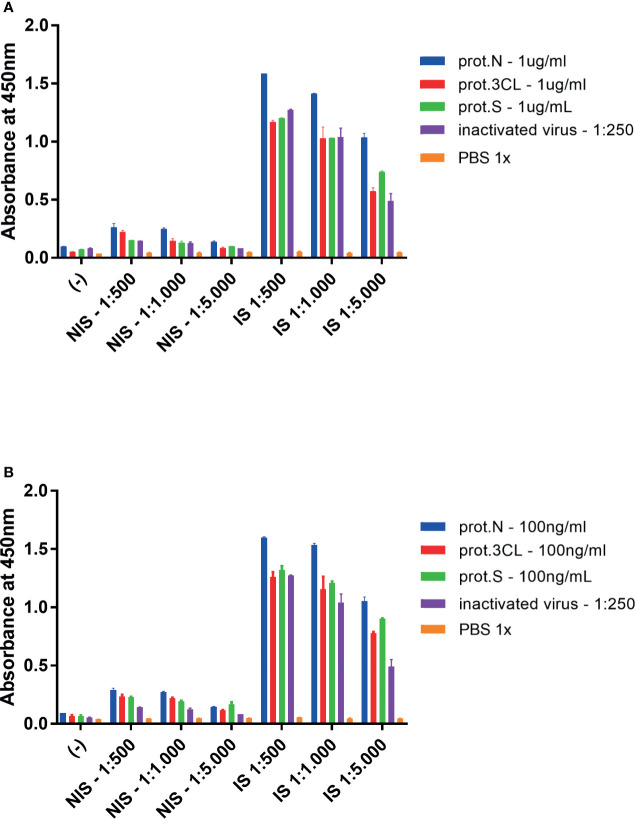
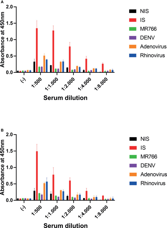
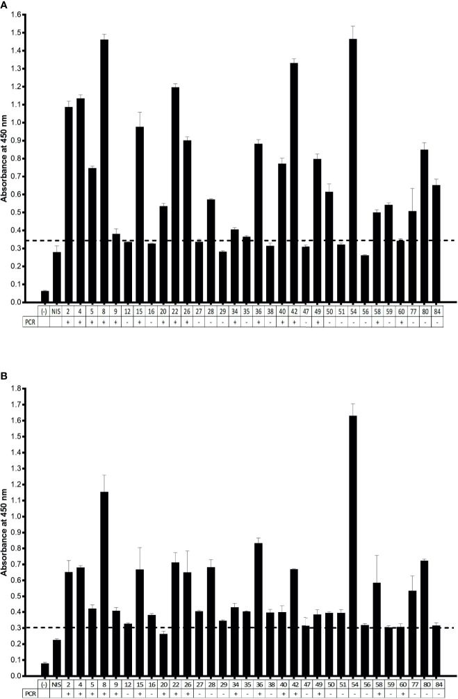
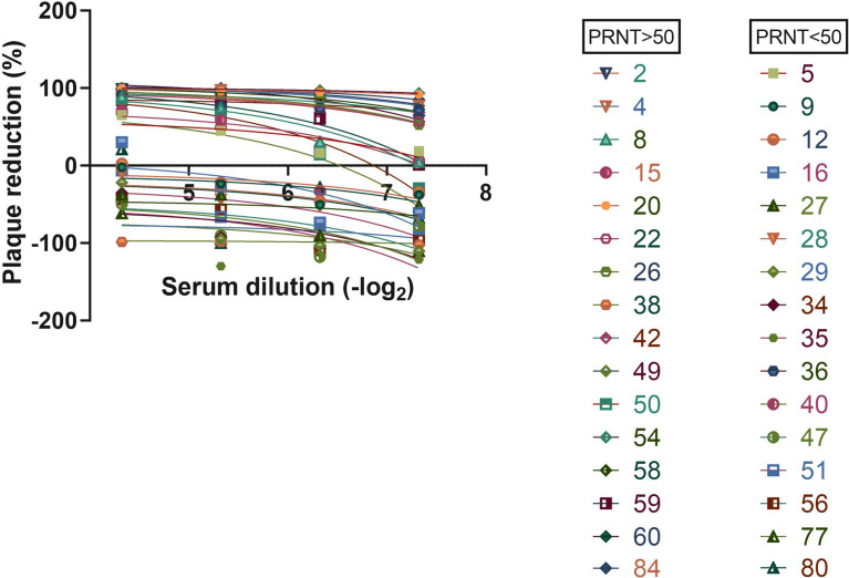
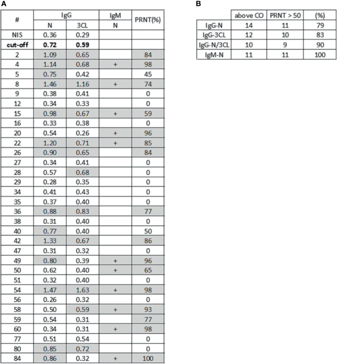
Serological testing is a powerful tool in epidemiological studies for understanding viral circulation and assessing the effectiveness of virus control measures, as is the case of SARS-CoV-2, the pathogenic agent of COVID-19. Immunoassays can quantitatively reveal the concentration of antiviral antibodies. The assessment of antiviral antibody titers may provide information on virus exposure, and changes in IgG levels are also indicative of a reduction in viral circulation. In this work, we describe a serological study for the evaluation of antiviral IgG and IgM antibodies and their correlation with antiviral activity. The serological assay for IgG detection used two SARS-CoV-2 proteins as antigens, the nucleocapsid N protein and the 3CL protease. Cross-reactivity tests in animals have shown high selectivity for detection of antiviral antibodies, using both the N and 3CL antigens. Using samples of human serum from individuals previously diagnosed by PCR for COVID-19, we observed high sensitivity of the ELISA assay. Serological results with human samples also suggest that the combination of higher titers of antiviral IgG antibodies to different antigen targets may be associated with greater neutralization activity, which can be enhanced in the presence of antiviral IgM antibodies.

Keywords: 3CL; COVID-19; SARS-CoV-2; immunoassay; nucleocapsid; seroneutralization.


Conflict of interest statement

The authors declare that the research was conducted in the absence of any commercial or financial relationships that could be construed as a potential conflict of interest.

Figures

Figure 1
Figure 1
ELISA assay for detection of mice antiviral antibodies. Mice were immunized with inactivated SARS-CoV-2 virus, and serum samples were collected with 14 days. (A) Plates were coated with 1.0 mg/ml of indicated viral antigens or 1:250 of inactivated virus. (B) plates were adsorbed with 100 ng/ml of indicated viral antigens or 1:250 of inactivated virus. Target antigens: prot.N (N protein), prot.3CL (3CL protein), prot.S (S protein), inactivated virus 1:250 (inactivated virus diluted 1:250), PBS 1x (1X PBS diluent as negative control). Two-way ANOVA, p < 0.05 for IS against NIS for all target antigens, but PBS 1x, in all dilutions. Representative experiment of two independent experiments. The serum of five animals was pooled for each condition.
Figure 2
Figure 2
Cross reactivity assay. Serum samples at indicated dilutions were incubated with antigen-coated plates. (A) Plates were coated with N protein. (B) Plates were coated with 3CL protein. (−) negative control, no serum; 1:500, 1:1,000, 1:2,000, 1:4,000, and 1:8,000: serum dilution; NIS, non-immune serum; IS,immune serum of mice immunized with inactivated SARS-CoV-2; MR766, immune serum of ZIKV challenged mice; DENV, immune serum of DENV challenged mice; Adenovirus, immune serum of Adenovirus challenged mice and Rhinovirus: immune serum of Rhinovirus challenged mice. Two-way ANOVA, p < 0.05 for IS against NIS in all dilutions, but 3CL 1:8000. The serum of five animals was pooled for each condition.
Figure 3
Figure 3
ELISA assay for IgG detection in human samples. (A) Serum samples diluted 1:100 were incubated to N protein-coated ELISA plates. (B) Serum samples diluted 1:100 were incubated to 3CL protein-coated ELISA plates. (−) negative control, absence of serum; NIS: non-immune human serum. the PCR status of nasal and oropharyngeal swab related to serum donors is indicated below the graph. Cutoff was set to NIS × 1.3. Representative experiment of two independent experiments.
Figure 4
Figure 4
Serum neutralization assay. Virus was incubated with indicated serum dilution following cell infection. Plaque reduction was calculated.
Figure 5
Figure 5
Serological profiling and virus neutralization. (A) The table shows absorbance signal for antiviral IgG reactive to N protein and 3CL protein by ELISA assay, IgM detection for N protein by LFA assay, and plaque reduction neutralization test (PRNT). Samples with absorbance signal above neutralization cutoff (twice NIS absorbance signal) for both antigens and samples with PRNT higher than 50% are highlighted in gray. (B) The table shows samples that exhibited high signal for antiviral antibodies to N protein (N), 3CL protease (3CL) and simultaneously (N and 3CL protein), in association to PRNT. In the “above CO” column, we have high IgG signal samples above the IgG positive cutoff (CO). The column PRNT>50 shows samples with neutralization activity higher than 50%. The last column shows the percentage of high neutralization samples that also have IgG signal above the cutoff, or IgM detected.

References

    1. Ruch TR, Machamer CE. The Coronavirus E Protein: Assembly and Beyond. Viruses (2012) 4(3):363–82. 10.3390/v4030363 - DOI - PMC - PubMed
    1. Satarker S, Nampoothiri M. Structural Proteins in Severe Acute Respiratory Syndrome Coronavirus-2. Arch Med Res (2020) 51(6):482–91. 10.1016/j.arcmed.2020.05.012 - DOI - PMC - PubMed
    1. Leung DT, Tam FC, Ma CH, Chan PK, Cheung JL, Niu H, et al. Antibody Response of Patients With Severe Acute Respiratory Syndrome (SARS) Targets the Viral Nucleocapsid. J Infect Dis (2004) 190(2):379–86. 10.1086/422040 - DOI - PMC - PubMed
    1. Wu A, Peng Y, Huang B, Ding X, Wang X, Niu P, et al. Genome Composition and Divergence of the Novel Coronavirus (2019-Ncov) Originating in China. Cell Host Microbe (2020) 27(3):325–8. 10.1016/j.chom.2020.02.001 - DOI - PMC - PubMed
    1. Liu Y, Liang C, Xin L, Ren X, Tian L, Ju X, et al. The Development of Coronavirus 3c-Like Protease (3CL(Pro)) Inhibitors From 2010 to 2020. Eur J Med Chem (2020) 206:112711. 10.1016/j.ejmech.2020.112711 - DOI - PMC - PubMed

Publication types

MeSH terms