. 2021 Aug 19:12:717370.
doi: 10.3389/fimmu.2021.717370. eCollection 2021.

Decreased USP2a Expression Inhibits Trophoblast Invasion and Associates With Recurrent Miscarriage

Affiliations

Decreased USP2a Expression Inhibits Trophoblast Invasion and Associates With Recurrent Miscarriage

Jiayu Wang et al. Front Immunol. .

Abstract

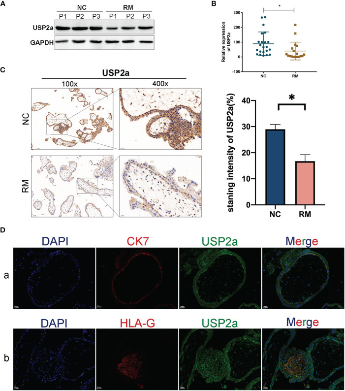
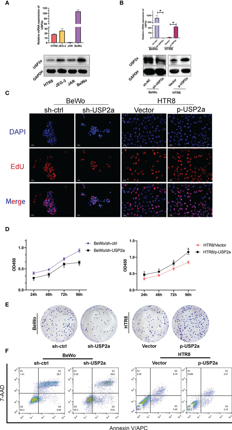
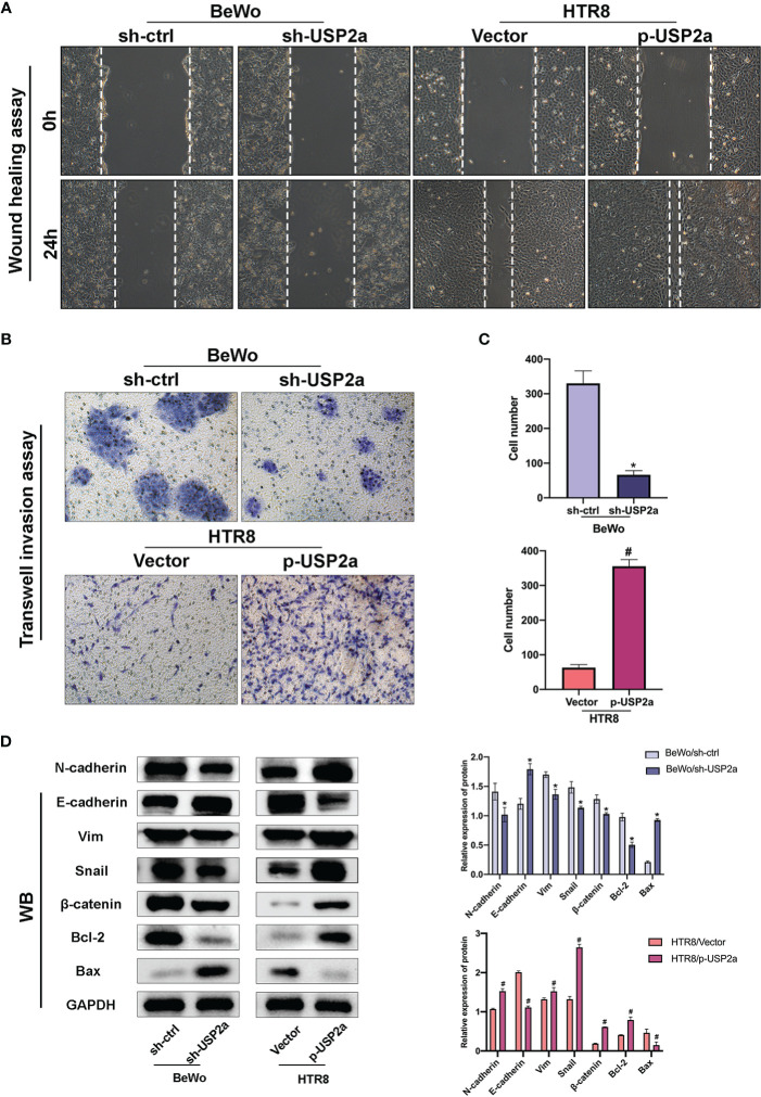
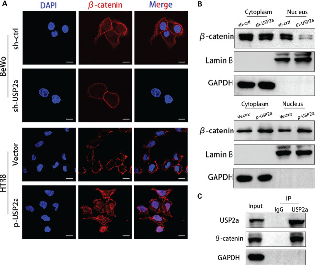
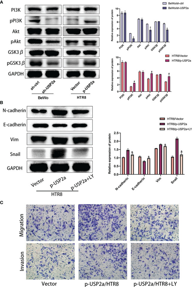
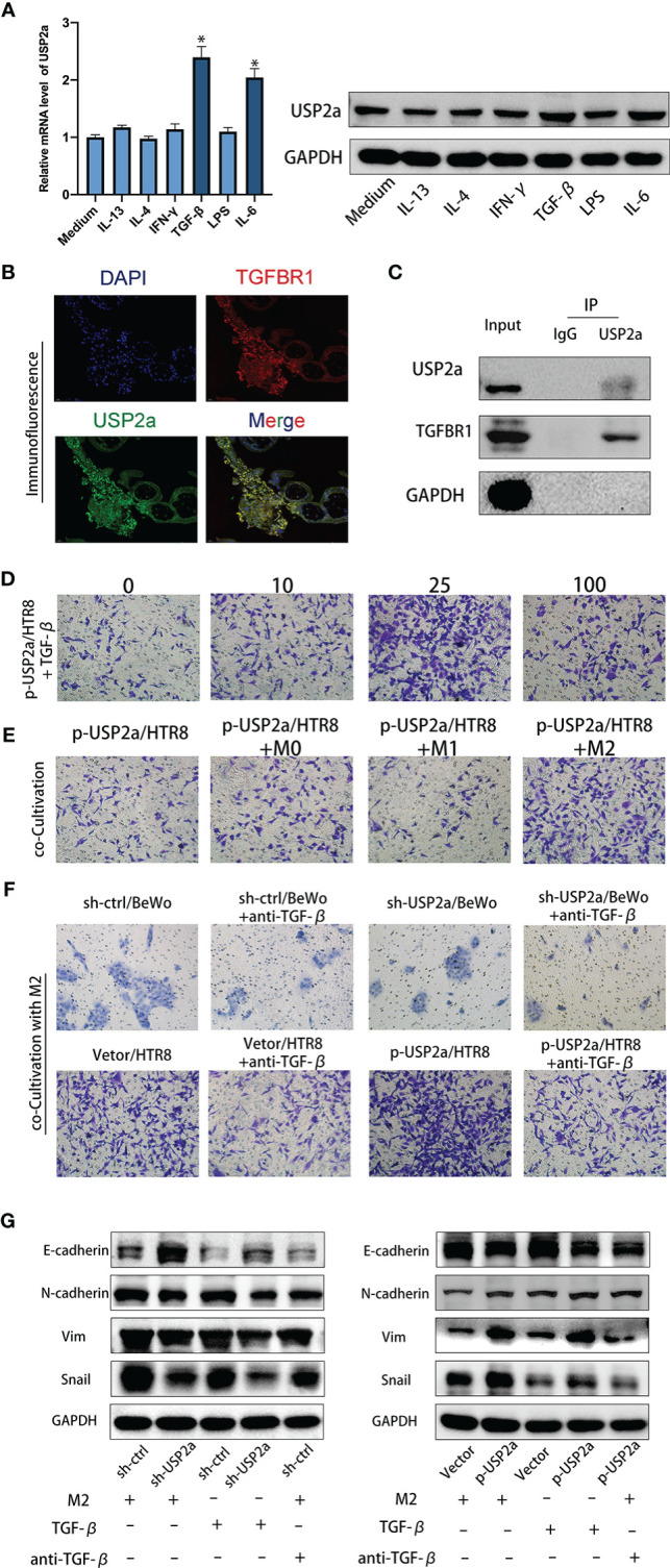
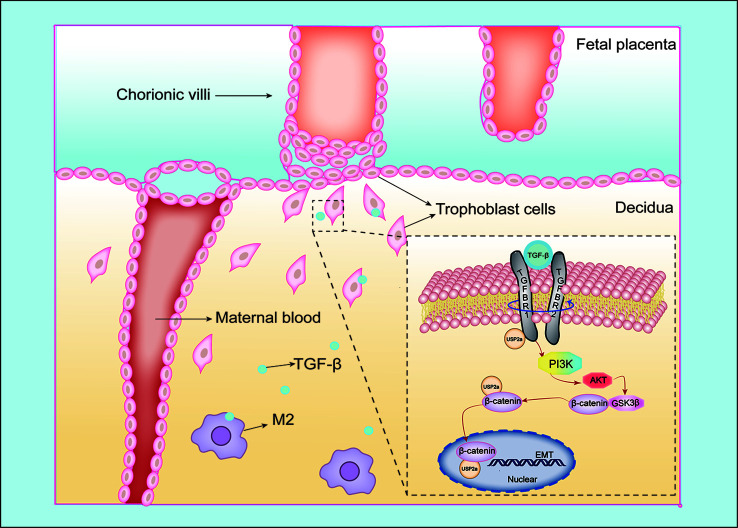
An appropriate development of the placenta consisting of trophoblast cell migration, invasion, proliferation, and apoptosis, is essential to establishing and maintaining a successful pregnancy. Ubiquitin-specific protease 2a (USP2a) regulates the processes of metastasis in multiple tumor cells. Yet, no known research has focused on exploring the effect of USP2a on trophoblasts and its possible mechanism in the pathogenies of recurrent miscarriage (RM). In this study, we first detected the decreased mRNA levels and the protein levels of USP2a in placental villous tissue samples from the RM patients. In vitro assays verified that overexpression of USP2a promoted human trophoblast proliferation, migration, invasion, whereas knockdown of USP2a inhibited these processes. Mechanistically, USP2a activated PI3K/Akt/GSK3β signaling pathway to promote nuclear translocation of β-catenin and further activated epithelial-mesenchymal transition (EMT) in the trophoblasts. Moreover, transforming growth factor-beta (TGF-β) up-regulated USP2a expression in trophoblasts. Interestingly, M2 macrophage secreted TGF-β induced trophoblast migration and invasion, and an anti-TGF-β antibody alleviated this effect. Collectively, this study indicated that USP2a regulated trophoblast invasion and that abnormal USP2a expression might lead to aberrant trophoblast invasion, thus contributing to RM.

Keywords: TGF-β; USP2a; decidual macrophage; recurrent miscarriage; trophoblast invasion.


Conflict of interest statement

The authors declare that the research was conducted in the absence of any commercial or financial relationships that could be construed as a potential conflict of interest.

Figures

Figure 1
Figure 1
USP2a is down-regulated in placental in patients with recurrent miscarriage (RM). (A, B) Comparison of relative mRNA and protein expression levels of USP2a in placental villous tissues between RM patients (n = 18) and normal women (n = 22) by RT‐PCR and western blot. (C) Representative immunohistochemical images of villous expression and localization of USP2a in RM patients (n = 22) and normal women (n = 25). The staining intensity was quantified using the ImageJ software. Graphs show the means ± SD. Magnification, 100× and 400×. (D) Representative immunofluorescence images: (a) double immune-staining of CK7 (red) and USP2a (green). Magnification, 400×; (b) double immune-staining of HLA-G (red) and USP2a (green) Magnification, 400×. *P < 0.05. Scale bar = 100 μm (100×); Scale bar = 20 μm (400×). mRNA, messenger RNA; RT‐PCR, quantitative real‐time polymerase chain reaction; RM, recurrent miscarriage; USP2a, ubiquitin‐specific protease 2a; CK7, cytokeratin 7; HLA-G, human leukocyte antigen G.
Figure 2
Figure 2
USP2a promoted human trophoblast‐derived cell lines proliferation and inhibited cell apoptosis in vitro. (A) Relative USP2a mRNA and protein expression levels in four human trophoblast‐derived cell lines were measured by RT‐PCR and western blot, respectively. GAPDH was used as the loading control. (B) Knockdown of USP2a by shRNA (sh‐USP2a) and upregulation of USP2a by plasmid expression were conducted in the BeWo and HTR8 cell lines. The mRNA expression and protein level of USP2a were confirmed by RT‐PCR and western blot. (C) Representative images of EdU labeling assay in p-USP2a/HTR8 and sh-USP2a/BeWo. DAPI staining blue; EdU staining red. Scale bar = 10 μm (D) Cell proliferation was measured 48 hours after transfection using the CCK8 assay. (E) Knocking down USP2a inhibits cell clone formation significantly in BeWo cells. Overexpression of USP2a promotes cell clone formation in HTR8 cells. (F) Quantitative flow cytometry measurements of apoptosis in trophoblast cell lines after transfection of either p-USP2a or shRNA. The percentage of live (7AAD−/Annexin V−), early apoptotic (7AAD−/Annexin V+), and dead (7AAD+/Annexin V+) cells was determined. *P < 0.05.
Figure 3
Figure 3
Effects of USP2a on the invasion and migration of trophoblast cells using the Transwell invasion assay and the wound-healing assay. (A) Wound healing scratches were imaged immediately and 24 h after initial scratch time to quantify relative migration. (B) After transfection, BeWo or HTR8 cells (in serum‐free medium) were seeded into inserts and incubated for 48 h for invasion assay. Cells at the lower surface of the membrane were counted and analyzed under a light microscope in five random fields. The staining intensity was quantified using the ImageJ software. Graphs show the means ± SD. (C) Western blot Graphs show the mean ± SD. Quantitative analysis of the average invasive cell numbers in each group. (D) The levels of E-cadherin, N-cadherin, Vimentin, Snail, β-catenin, Bax, and Bcl2 were measured using western blotting and quantitative analysis of the proteins was performed. *P < 0.05 vs. sh-Ctrl # P < 0.05 vs control vector, original magnification: ×100 (A) ×200 (B).
Figure 4
Figure 4
Reduced USP2a Expression impairs nucleocytoplasmic translocation of β-catenin. Knockdown of USP2a attenuates nuclear translocation of β-catenin in BeWo cells. Overexpression of USP2a induces nuclear translocation of β-catenin in HTR8 cells BeWo cells were transfected with sh-Ctrl or sh-USP2a, and HTR8 cells were transfected with p-USP2a or vector. Both cells were cultured for 48 hours. (A) The cells were seeded to immunofluorescent staining for β-catenin (red) and counterstained with DAPI (blue) to assess the subcellular localization of β-catenin and (B) western blot was performed to compare the levels of β-catenin in the nucleus and cytoplasm. GAPDH and lamin B were used as the loading control. (C) Co-IP assays of the interaction between USP2a and β-catenin in HTR8. Scale bar = 10μm.
Figure 5
Figure 5
USP2a promotes EMT, migration and invasion of trophoblasts via activating PI3K/Akt/GSK3β pathway. (A) Western blot analyzes relative protein level of sh-Ctrl/BeWo, sh-USP2a/BeWo, Vector/HTR8, p-USP2a/HTR8. (B) Western blot analysis of sh-Ctrl/BeWo, sh-USP2a/BeWo, Vector/HTR-8, p-USP2a/HTR8 in the presence or absence of LY29004 (20 μM). (C) Migration and invasion capacity in sh-Ctrl/BeWo, sh-USP2a/BeWo, Vector/HTR8, p-USP2a/HTR8 in the presence or absence of LY294002 (20 μM) was determined by transwell system. Representative photographs of migratory and invasive cells (magnification, ×200) are shown. *P < 0.05, vs. sh-Ctrl # P < 0.05 vs vector. & P < 0.05 vs p-USP2a/HTR8.
Figure 6
Figure 6
M2 Macrophage-secreted TGF-β cooperate with USP2a in trophoblasts to promote invasion. (A) Western blot and quantitative RT-PCR analysis of USP2a expression in HTR8 treated with IFN-γ, IL-13, IL-4, IL-6, LPS, or TGF-β for 24 hours. (B) Immunostaining for TGFBR1 and USP2a in villous from normal pregnancy woman (Scale bar = 20 μm). (C) co-IP assays of the interaction between USP2a and TGFBR1 in p-USP2a/HTR8. (D) Cell invasion capacity in HTR8 alone, HTR8 with 10ng/ml, 25ng/ml, or 100ng/ml rhTGF-β by transwell system. (E) Cell invasion capacity in p-USP2a/HTR8 alone, p-USP2a/HTR8 co-cultured with M0 macrophage, co-cultured with M1 macrophage, and co-cultured with M2 macrophage by transwell system. (F) Cell invasion capacity in sh-Ctrl/BeWo, sh-USP2a/BeWo, Vector/HTR8, p-USP2a/HTR8 co-cultured with M2 macrophage or above cells co-cultured with TGF-β depleted M2 macrophage were determined by transwell system, respectively. (G) Western blot assay of EMT proteins in sh-Ctrl/BeWo, sh-USP2a/BeWo, Vector/HTR8, p-USP2a/HTR8 co-cultured with M2 macrophage or above cells co-cultured with TGF-β depleted M2 macrophage were determined by transwell system, respectively. *P < 0.05, vs. HTR8. DAPI staining blue; TGFBR1 staining red; USP2a staining green. Representative photographs of invasive cells (magnification, ×200) are shown.
Figure 7
Figure 7
Schematic diagram of the interaction between trophoblasts and macrophages at the maternal-fetal interface. Our study illustrated USP2a facilitates the EMT programme to enhance migration and invasion of trophoblasts secreted by phosphorylating PI3K/Akt/GSK3β/β-catenin signaling pathways. In the meantime, USP2a could interreact with β-catenin as a “shortcut” to promote nuclear β-catenin accumulation. TGF-β secreted by M2 macrophage could work together with USP2a to promote trophoblast invasion. Thereby, USP2a participates in regulating the establishment and maintenance of normal pregnancy.

Similar articles

Cited by

References

    1. Rai R, Regan L. Recurrent Miscarriage. Lancet (2006) 368(9535):601–11. 10.1016/S0140-6736(06)69204-0 - DOI - PubMed
    1. Popescu F, Jaslow CR, Kutteh WH. Recurrent Pregnancy Loss Evaluation Combined With 24-Chromosome Microarray of Miscarriage Tissue Provides a Probable or Definite Cause of Pregnancy Loss in Over 90% of Patients. Hum Reprod (2018) 33(4):579–87. 10.1093/humrep/dey021 - DOI - PubMed
    1. Arck PC, Hecher K. Fetomaternal Immune Cross-Talk and Its Consequences for Maternal and Offspring’s Health. Nat Med (2013) 19(5):548–56. 10.1038/nm.3160 - DOI - PubMed
    1. Mayhew TM. Turnover of Human Villous Trophoblast in Normal Pregnancy: What do We Know and What do We Need to Know? Placenta (2014) 35(4):229–40. 10.1016/j.placenta.2014.01.011 - DOI - PubMed
    1. Nieto MA. Epithelial-Mesenchymal Transitions in Development and Disease: Old Views and New Perspectives. Int J Dev Biol (2009) 53(8-10):1541–7. 10.1387/ijdb.072410mn - DOI - PubMed

Publication types

MeSH terms