. 2021 Aug 19:11:700700.
doi: 10.3389/fonc.2021.700700. eCollection 2021.

Identification of PAFAH1B3 as Candidate Prognosis Marker and Potential Therapeutic Target for Hepatocellular Carcinoma

Affiliations

Identification of PAFAH1B3 as Candidate Prognosis Marker and Potential Therapeutic Target for Hepatocellular Carcinoma

Weikang Xu et al. Front Oncol. .

Abstract

Background: Hepatocellular carcinoma (HCC) is the fourth leading cause of cancer-related deaths worldwide. PAFAH1B3 plays an important role on occurrence and development in a variety tumor. However, the function of PAFAH1B3 in HCC remains unclear.

Methods: The TIMER, ONCOMINE, Human Protein Atlas (HPA), GEPIA, The Cancer Genome Atlas (TCGA), HCCDB, UALCAN and LinkedOmics database were used to analyze the prognostic value, co-expression genes and regulator networks of PAFAH1B3 in HCC. siRNA transfections and inhibitor of PAFAH1B3 P11 were used to verify the anti-tumor effect on HCC cell lines. Gene expression was detected by qRT-PCR. The functions of PAFAH1B3 downregulation in HCC cell lines were investigated using cell cycle analysis, apoptosis detection, CCK8 assay and transwell assay. Western blot was used to evaluate the role of PAFAH1B3 on metabolic pathways in HCC cells.

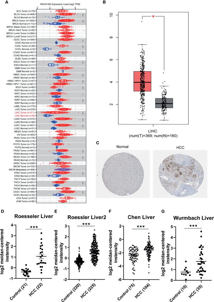
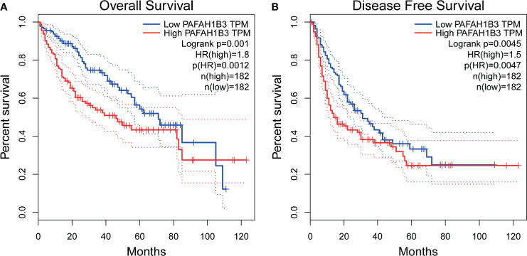
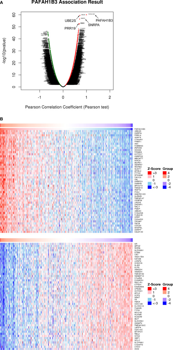
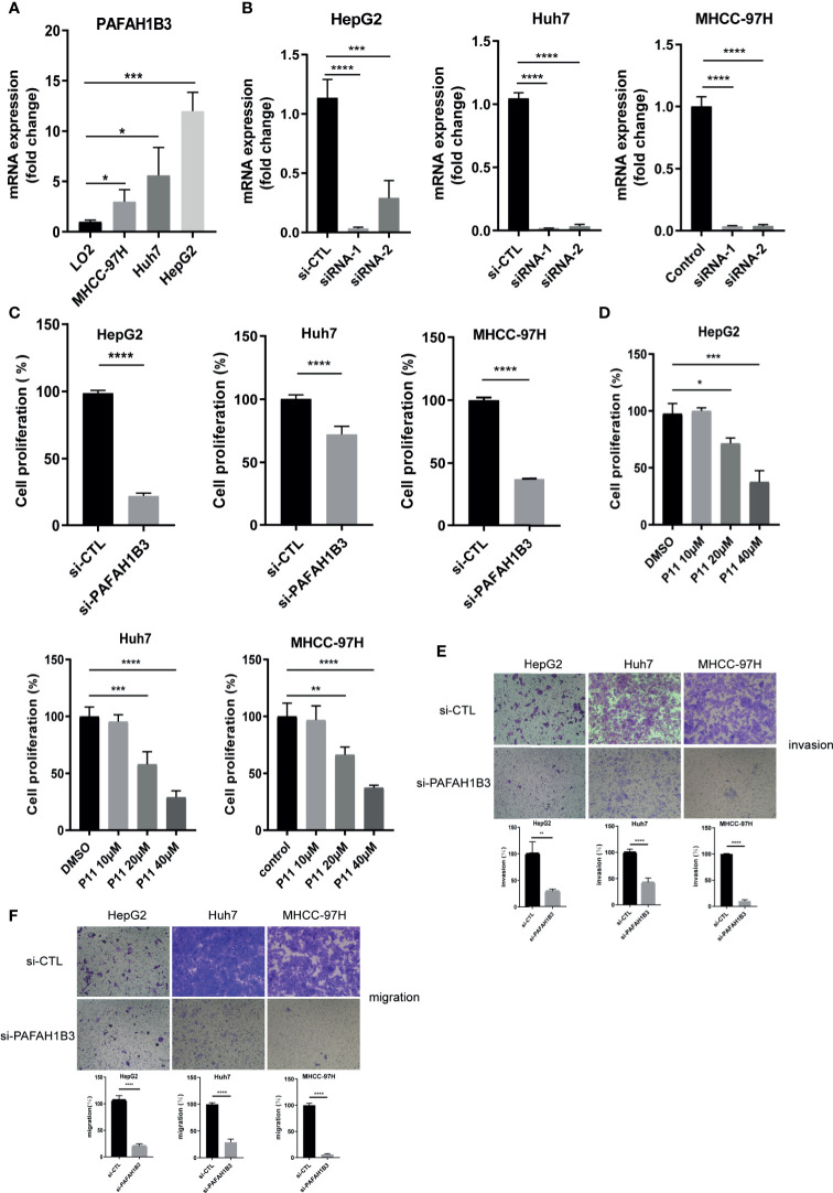
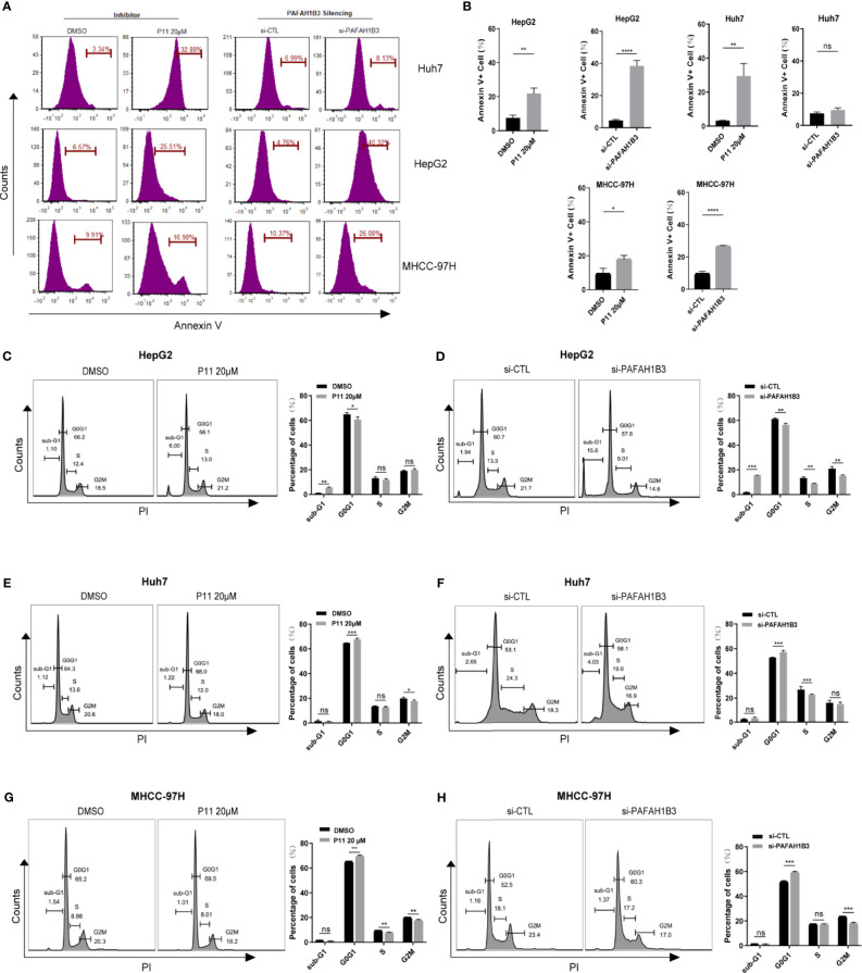
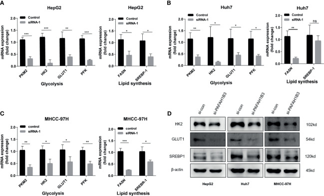
Results: Based on the data from databases, the expression of PAFAH1B3 was remarkably increased in HCC patients. High expression of PAFAH1B3 was associated with poorer overall survival (OS) and disease-free survival (DFS). And PAFAH1B3 was notably linked to age, sex, grade, stage, race, and TP53 mutational status. Then, the functional network analysis showed PAFAH1B3 may be involved in HCC through cell cycle, cell metabolism, spliceosome, and RNA transport. Furthermore, the mRNA expression of PAFAH1B3 was also increased in HCC cell lines. Flow cytometry analysis showed that PAFAH1B3 manipulated apoptosis and cell cycle regulation. CCK8 assay showed that PAFAH1B3 silencing or pharmacologic inhibitor of PAFAH1B3 inhibited the proliferation of HepG2, Huh7 and MHCC-97H cells. Transwell assay results showed that PAFAH1B3 silencing also significantly impaired the invasion and migratory ability of HCC cells. In addition, PAFAH1B3 silencing significantly downregulated the expression of glycolysis and lipid synthesis signaling pathways.

Conclusion: Our findings suggested that PAFAH1B3 plays a critical role in progression of HCC. PAFAH1B3 as a prognosis marker and potential target for HCC has prospective clinical significance.

Keywords: PAFAH1B3; biomarker; cancer databases; hepatocellular carcinoma; prognosis.


Conflict of interest statement

The authors declare that the research was conducted in the absence of any commercial or financial relationships that could be construed as a potential conflict of interest.

Figures

Figure 1
Figure 1
Expression of PAFAH1B3 in hepatocellular carcinoma (HCC). (A) The transcription level of PAFAH1B3 in TIMER databases. (B) The transcription level of PAFAH1B3 in ONCOMINE databases. (C) The protein expression of PAFAH1B3 in LIHC (HPA). (D) Boxplot showing the levels of PAFAH1B3 mRNA in the Roessler Liver. (E) Boxplot showing the levels of PAFAH1B3 mRNA in the Roessler Liver 2 datasets. (F) Boxplot showing the levels of PAFAH1B3 mRNA in the Chen Liver datasets. (G). Boxplot showing the levels of PAFAH1B3 mRNA in the Wurmbach Liver datasets. *p < 0.05; ***p < 0.001.
Figure 2
Figure 2
Prognostic Value of PAFAH1B3 in Patients with hepatocellular carcinoma (HCC) (GEPIA). (A) Overall survival (OS) of PAFAH1B3 in HCC. (B) Disease-free survival (DFS) of PAFAH1B3 in HCC.
Figure 3
Figure 3
Coexpression of PAFAH1B3 in hepatocellular carcinoma (HCC) (LinkedOmics). (A) Total genes highly correlated with PAFAH1B3. (B) Heatmaps showing the 50 significant genes positively and negatively correlated with PAFAH1B3 in LIHC. Red and blue indicate positively and negatively correlated genes, respectively.
Figure 4
Figure 4
High expression of PAFAH1B3 related to the proliferation and metastasis of HCC cells. (A) Levels of PAFAH1B3 mRNA in LO2, MHCC-97H, Huh7, and HepG2 cell lines. (B) Quantitation of the levels of PAFAH1B3 mRNA in HepG2, Huh7 and MHCC-97H cells by qRT-PCR after transfection for 48 h with different siRNAs targeting PAFAH1B3 (si-1 and si-2) or a control siRNA. (C) Proliferation of HepG2 and Huh7 cells after silencing PAFAH1B3 for 48 h (D) Proliferation of HepG2 and Huh7 cells after pharmacologic inhibitor of PAFAH1B3 for 48 h (E) Invasion assay. (F) Migratory assay. *p < 0.05; **p < 0.01; ***p < 0.001; ****p < 0.0001.
Figure 5
Figure 5
The effects of PAFAH1B3 on HCC cell apoptosis and cell cycle. (A, B) The flow cytometry analysis revealed the effects of PAFAH1B3 silencing or inhibitor P11 on cell apoptosis. (C–H) Cell cycle analysis in HepG2, Huh7 and MHCC-97H cells. *p < 0.05; **p < 0.01; ***p < 0.001; ****p < 0.0001. NS, no significance.
Figure 6
Figure 6
PAFAH1B3 downregulates glycolysis and lipid synthesis signaling pathways. (A–C) Analysis of glycolysis-and lipid synthesis-related genes (PKM2, HK2, GLUT1, PFK, FASN and SREBP-1) in HepG2 Huh7 and MHCC-97H cells after silencing PAFAH1B3 for 48 h. (D) Analysis of glycolysis-related key enzymes and lipid synthesis-related transcription factor (HK2, GLUT1 and SREBP-1) in HCC cells by Western blot after silencing PAFAH1B3 for 48 h. *p < 0.05; **p < 0.01; ***p < 0.001. NS, no significance.

Similar articles

Cited by

References

    1. Bray F, Ferlay J, Soerjomataram I, Siegel RL, Torre LA, Jemal A. Global Cancer Statistics 2018: GLOBOCAN Estimates of Incidence and Mortality Worldwide for 36 Cancers in 185 Countries. CA Cancer J Clin (2018) 68(6):394–424. 10.3322/caac.21492 - DOI - PubMed
    1. Forner A, Reig M, Bruix J. Hepatocellular Carcinoma. Lancet (2018) 391(10127):1301–14. 10.1016/s0140-6736(18)30010-2 - DOI - PubMed
    1. McGlynn KA, Petrick JL, El-Serag HB. Epidemiology of Hepatocellular Carcinoma. Hepatology (2021) 73 Suppl 1(Suppl 1):4–13. 10.1002/hep.31288 - DOI - PMC - PubMed
    1. Karin M. New Insights Into the Pathogenesis and Treatment of non-Viral Hepatocellular Carcinoma: A Balancing Act Between Immunosuppression and Immunosurveillance. Precis Clin Med (2018) 1(1):21–8. 10.1093/pcmedi/pby005 - DOI - PMC - PubMed
    1. Nault JC, Villanueva A. Biomarkers for Hepatobiliary Cancers. Hepatology (2021) 73 Suppl 1:115–27. 10.1002/hep.31175 - DOI - PubMed