Three-Dimensional Kinematic Motion of the Craniocervical Junction of Chihuahuas and Labrador Retrievers
- PMID: 34490400
- PMCID: PMC8417724
- DOI: 10.3389/fvets.2021.709967
Three-Dimensional Kinematic Motion of the Craniocervical Junction of Chihuahuas and Labrador Retrievers
Abstract
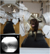
All vertebrate species have a distinct morphology and movement pattern, which reflect the adaption of the animal to its habitat. Yet, our knowledge of motion patterns of the craniocervical junction of dogs is very limited. The aim of this prospective study is to perform a detailed analysis and description of three-dimensional craniocervical motion during locomotion in clinically sound Chihuahuas and Labrador retrievers. This study presents the first in vivo recorded motions of the craniocervical junction of clinically sound Chihuahuas (n = 8) and clinically sound Labrador retrievers (n = 3) using biplanar fluoroscopy. Scientific rotoscoping was used to reconstruct three-dimensional kinematics during locomotion. The same basic motion patterns were found in Chihuahuas and Labrador retrievers during walking. Sagittal, lateral, and axial rotation could be observed in both the atlantoaxial and the atlantooccipital joints during head motion and locomotion. Lateral and axial rotation occurred as a coupled motion pattern. The amplitudes of axial and lateral rotation of the total upper cervical motion and the atlantoaxial joint were higher in Labrador retrievers than in Chihuahuas. The range of motion (ROM) maxima were 20°, 26°, and 24° in the sagittal, lateral, and axial planes, respectively, of the atlantoaxial joint. ROM maxima of 30°, 16°, and 18° in the sagittal, lateral, and axial planes, respectively, were found at the atlantooccipital joint. The average absolute sagittal rotation of the atlas was slightly higher in Chihuahuas (between 9.1 ± 6.8° and 18.7 ± 9.9°) as compared with that of Labrador retrievers (between 5.7 ± 4.6° and 14.5 ± 2.6°), which corresponds to the more acute angle of the atlas in Chihuahuas. Individual differences for example, varying in amplitude or time of occurrence are reported.
Keywords: cervical spine; craniocervical motion; dog locomotion; scientific rotoscoping; three-dimensional kinematics.
Copyright © 2021 Schikowski, Eley, Kelleners, Schmidt and Fischer.
Conflict of interest statement
The authors declare that the research was conducted in the absence of any commercial or financial relationships that could be construed as a potential conflict of interest.
Figures


References
-
- Arnold P. Evolution of the mammalian neck from developmental, morpho-functional, and paleontological perspectives. J Mamm Evol. (2020) 28:1–11. 10.1007/s10914-020-09506-9 - DOI
-
- McLear RC, Saunders HM. Atlantoaxial mobility in the dog. Vet Radiol Ultrasound. (2000) 41:558.
-
- Morgan J, Miyabayashi T, Choy S. Cervical spine motion: radiographic study. Am J Vet Res. (1986) 47:2165–9. - PubMed
LinkOut - more resources
Full Text Sources

