Review
. 2021 Sep 15;101(9):adv00548.
doi: 10.2340/00015555-3917.

Dermoscopic Evaluation of Inflammatory Nail Disorders and Their Mimics

Affiliations
Review

Dermoscopic Evaluation of Inflammatory Nail Disorders and Their Mimics

Sophie Soyeon Lim et al. Acta Derm Venereol. .

Abstract

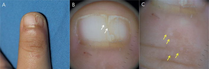
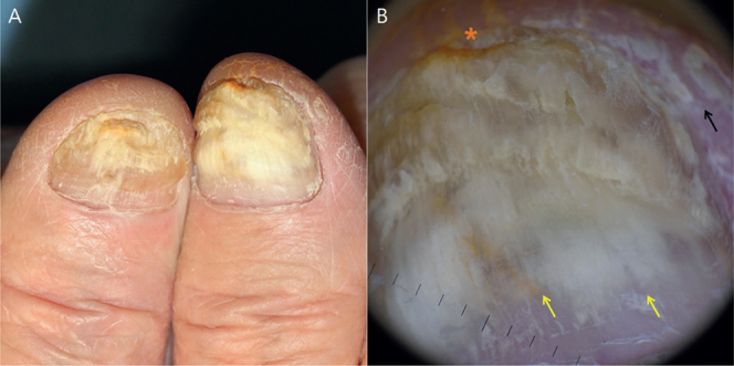
Nail dermoscopy (onychoscopy) is a valuable diagnostic tool for evaluating diseases in the nail apparatus. It is non-invasive, allowing clinicians to prioritize particular nails for biopsy. Thus, it can improve diagnostic accuracy and expedite treatment. Evaluating inflammatory nail disorders using onychoscopy is a relatively new approach to clinical assessment and has the potential to augment clinical care. This review highlights key dermoscopic features of major inflammatory nail disorders, including trachyonychia, nail psoriasis, nail lichen planus, onychotillomania, nail lichen striatus and allergic contact dermatitis due to artificial nails. It also illustrates their management and differential diagnoses, including onychomycosis, onycholysis, nail dystrophy due to systemic amyloidosis and malignant nail tumours. Limitations of this review included the low amount of literature on this topic and non-standardized terminology used among research-ers. As onychoscopy is a relatively new technique, further studies and standardization of terminology are warranted to consolidate the role of dermoscopy in evaluating inflammatory nail disorders.

Keywords: allergic contact dermatitis; lichen planus; lichen striatus; onychotillomania; psoriasis; trachyonychia.


Conflict of interest statement

The authors have no conflicts of interest to declare.

Figures

Fig. 1
Fig. 1
Trachyonychia. (A) and (C) Clinical photography demonstrates the “sandpaper-like” nail surface in idiopathic trachyonychia. (B) and (D) Dermoscopy shows involvement of >50% of the proximal nail plate width (blue arrow), proximal nail involvement, longitudinal ridging (white arrows), splinter haemorrhages (red arrow), opaque lunula (white asterisk), and thick and ragged cuticles (green arrows).
Fig. 2
Fig. 2
Nail psoriasis. (A) Clinical photography shows nail changes and erythematous scaly papules/plaques on the dorsum of fingers. (B) Dermoscopy of nail psoriasis highlights the pitting (white arrows), splinter haemorrhages (yellow arrows), distal onycholysis (blue arrow) and oil drop sign/salmon patch (red asterisk).
Fig. 3
Fig. 3
Nail lichen planus. (A) Clinical photograph of nail lichen planus with atrophic and scarring changes. (B) Dermoscopy of nail lichen planus demonstrates onychorrhexis (yellow arrows), distal nail splitting (black arrow) and red chromonychia (red asterisk).
Fig. 4
Fig. 4
Onychotillomania. (A) Clinical photograph of onychotillomania. (B) Dermoscopy demonstrates scales (white arrows), loss of a nail plate (white asterisk), wavy lines (yellow arrows), haemorrhage (red arrows), and crust (black asterisk).
Fig. 5
Fig. 5
Nail lichen striatus. (A) Clinical photograph of nail lichen striatus. (B, C) Dermoscopic image shows fissuring of the nail plate (white arrows) and white papules on the skin in association with nail changes (yellow arrows).
Fig. 6
Fig. 6
Onychomycosis. (A) Clinical photography of onychomycosis shows the yellow, brittle nails. (B) Dermoscopy displays yellow discolouration of nail plate in the distal two-thirds of the nail plate, yellow/white streaks (yellow arrows), jagged proximal margin, subungual hyperkeratosis (orange asterisk) and periungual scales (black arrow).
Fig. 7
Fig. 7
Amelanotic nail melanoma. Dermoscopic image shows amelanotic nail apparatus melanoma with black dots, micro-haemorrhages, subungual hyperkeratosis, and partial nail dystrophy. (Inset: clinical photograph).

References

    1. Jacobsen AA, Tosti A. Trachyonychia and twenty-nail dystrophy: a comprehensive review and discussion of diagnostic accuracy. Skin Appendage Disord 2016; 2: 7–13. - PMC - PubMed
    1. Alkiewicz J. Trachyonychia. Ann Dermatol Venereol 1950; 10: 136–140.
    1. Starace M, Alessandrini A, Bruni F, Piraccini BM. Trachyonychia: a retrospective study of 122 patients in a period of 30 years. J Eur Acad Dermatol Venereol 2020; 34: 880–884. - PubMed
    1. Haber JS, Chairatchaneeboon M, Rubin AI. Trachyonychia: review and update on clinical aspects, histology, and therapy. Skin Appendage Disord 2016; 2: 109–115. - PMC - PubMed
    1. Tosti A, Fanti PA, Morelli R, Bardazzi F. Trachyonychia associated with alopecia areata: a clinical and pathologic study. J Am Acad Dermatol 1991; 25: 266–270. - PubMed