. 2022 Feb;75(2):353-368.
doi: 10.1002/hep.32140. Epub 2021 Dec 15.

Ductular reaction promotes intrahepatic angiogenesis through Slit2-Roundabout 1 signaling

Affiliations

Ductular reaction promotes intrahepatic angiogenesis through Slit2-Roundabout 1 signaling

Mar Coll et al. Hepatology. 2022 Feb.

Abstract

Background and aims: Ductular reaction (DR) expands in chronic liver diseases and correlates with disease severity. Besides its potential role in liver regeneration, DR plays a role in the wound-healing response of the liver, promoting periductular fibrosis and inflammatory cell recruitment. However, there is no information regarding its role in intrahepatic angiogenesis. In the current study we investigated the potential contribution of DR cells to hepatic vascular remodeling during chronic liver disease.

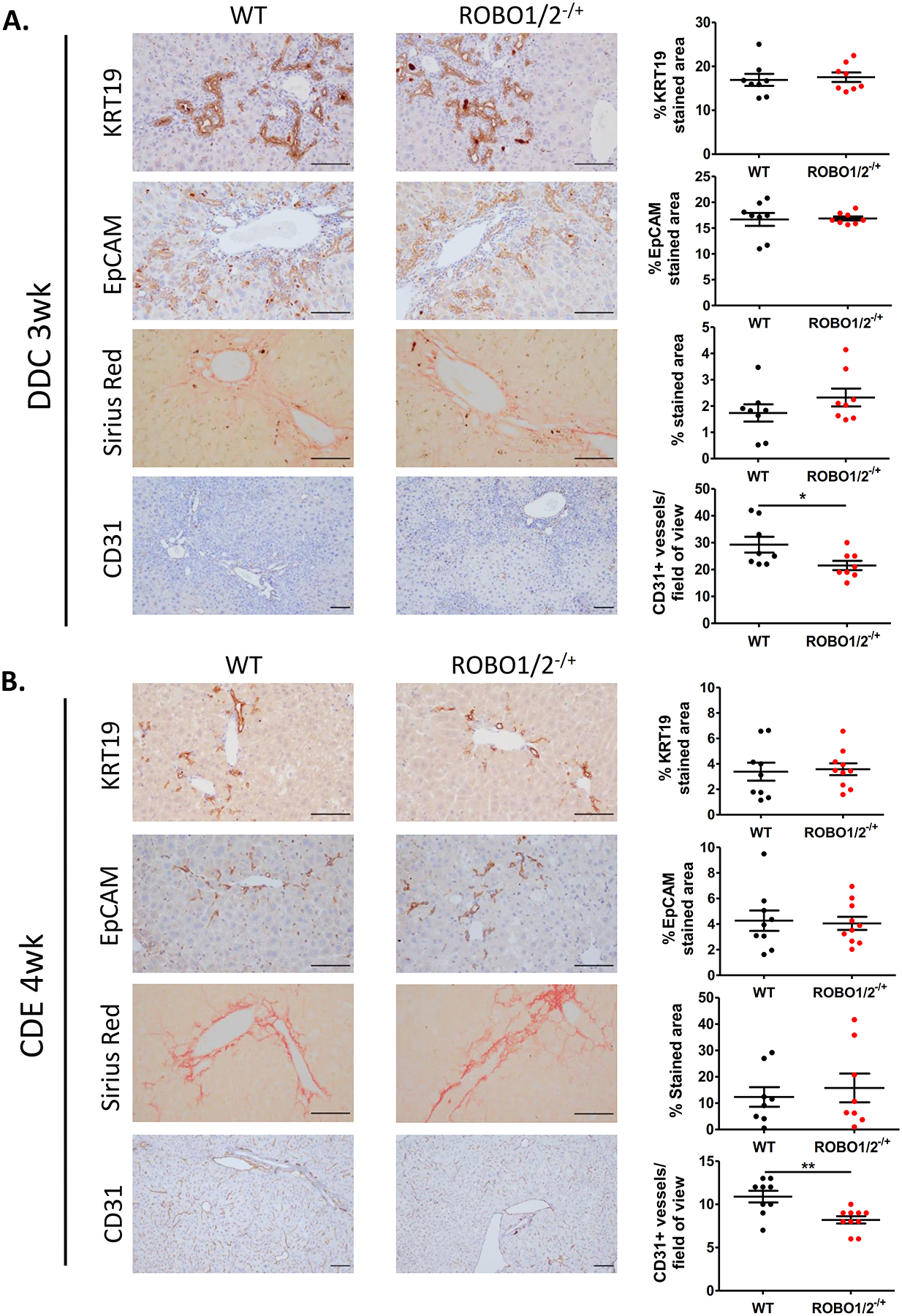
Approach and results: In mouse models of liver injury, DR cells express genes involved in angiogenesis. Among angiogenesis-related genes, the expression of Slit2 and its receptor Roundabout 1 (Robo1) was localized in DR cells and neoangiogenic vessels, respectively. The angiogenic role of the Slit2-Robo1 pathway in chronic liver disease was confirmed in ROBO1/2-/+ mice treated with 3,5-diethoxycarbonyl-1,4-dihydrocollidine, which displayed reduced intrahepatic neovascular density compared to wild-type mice. However, ROBO1/2 deficiency did not affect angiogenesis in partial hepatectomy. In patients with advanced alcohol-associated disease, angiogenesis was associated with DR, and up-regulation of SLIT2-ROBO1 correlated with DR and disease severity. In vitro, human liver-derived organoids produced SLIT2 and induced tube formation of endothelial cells.

Conclusions: Overall, our data indicate that DR expansion promotes angiogenesis through the Slit2-Robo1 pathway and recognize DR cells as key players in the liver wound-healing response.


Conflict of interest statement

CONFLICT OF INTEREST: Nothing to declare.

Figures

Figure 1.
Figure 1.. Gene expression and gene ontology (GO) analysis of a transcriptomic dataset derived from ductular reaction cells isolated from HNF1βCreERYFP and control mice.
(A) GO circle plot indicating the top 5 enriched biological processes related to ductular reaction cells of 3,5-diethoxycarbonyl-1,4-dihydro-collidin (DDC) and choline-deficient, ethionine-supplemented diet (CDE) models. The outer ring illustrates the expression sign (blue, down-regulated and red, up-regulated) of the genes included in each GO term. The color intensity of the inner ring shows the significance measured by the z-score for each category. (B) GOChord plot showing genes linked to their assigned GO terms. (C) Heat map displaying the top 75 significantly deregulated genes in YFP+ cells isolated from mice receiving DDC or CDE diets for 3 weeks compared to YFP+ cells of the control mice.
Figure 2.
Figure 2.. Gene and protein expression of Slit2-Robo1 signaling in mouse models involving ductular reaction proliferation.
(A) Gene expression analysis by qPCR of Slit2, Robo1, Robo4 and DR markers (Krt19 and Epcam) in whole liver tissue isolated from mice receiving a 3,5-diethoxycarbonyl-1,4-dihydro-collidin (DDC) (n=5) and choline-deficient, ethionine-supplemented diet (CDE) for 3 weeks (n=5). Expression values are represented as Log2 Fold change versus control mice. (B) Levels of gene expression measured by qPCR of key DR markers (Epcam and Krt19) and Slit2 and its receptor Robo1 in YFP+ cells and hepatocyte fraction from livers of mice receiving DDC for 3 weeks. Expression values are represented as fold change versus the expression in whole liver tissue. (C) Representative images at 10x of SLIT2 and ROBO1 immunohistochemistry in liver tissues of a mouse receiving DDC for 3 weeks show the expression of SLIT2 by DR cells and ROBO1 expression restricted to new vessels. Immunofluorescence of KRT19 (green) and SLIT2 (red) confirms co-localization of SLIT2 within the DR. Nuclei counterstaining was performed with DAPI (blue). Scale bar = 100μm. Data presented as mean ± SEM. *p<0.05, **P<0.01 and ***p<0.001.
Figure 3.
Figure 3.. Slit2-Robo1 signaling mediates angiogenesis in both DDC and CDE mouse models.
Representative immunohistochemical staining of KRT19, EPCAM, Sirius Red and CD31 staining in ROBO1/2−/+ and WT mice receiving (A) DDC diet for 3 weeks (n=8 in both groups) or (B) CDE diet for 4 weeks (n=9 in WT group, n=10 in ROBO1/2−/+ group). In both injury models, DR/progenitor cell expansion and fibrogenesis were evaluated by quantification of positive staining areas for KRT19/EPCAM and Sirius Red, respectively, measured by ImageJ Software. Neo-angiogenesis was quantified by counting the number of CD31 positive vessels surrounding the peri-portal areas/field of view. Scale bars = 100μm. Data is presented as mean ± SEM. Groups were compared by t-test analysis. *p<0.05 and **P<0.01.
Figure 4.
Figure 4.. Slit2-Robo1 signaling does not participate in partial hepatectomy regeneration.
(A) Gene expression analysis of liver progenitor cell markers (Krt19, Epcam and Sox9), Slit2 and Robo1 in mouse livers at early (24, 48 and 72 hours after surgery) and late (day 7 and day 28) time points after two thirds partial hepatectomy in WT mice (n= 3 to 5 mice per group). Fold change is represented as expression at each time point versus expression at day 0. Grouped analysis was compared by two-way ANOVA with Bonferroni post-test correction. *p<0.05, **p<0.01, ***p<0.001. (B) Two thirds partial hepatectomy was evaluated in WT and ROBO1/2−/+ mice (n= 9 in WT group and n=10 in ROBO1/2−/+ group), being sacrificed at 48 hours after the surgery. (C) Two thirds partial hepatectomy was performed in WT and ROBO1/2−/+ mice (n= 8 in WT group and n=7 in ROBO1/2−/+ group), and animals were sacrificed at day 7 after the surgery. (B) Representative images of cell proliferation at 48 hours evaluated by KI67 staining. (B-C) Representative immunohistochemical staining for CD31 and KRT19 in livers after 48 hours and 7 days after partial hepatectomy. DR expansion and cell proliferation were measured by the quantification of positive staining for KRT19 and KI67, respectively, by ImageJ Software. New vessel formation was evaluated by counting CD31+ vessels/field of view. Scale bars = 100μm. Data presented as mean ± SEM. Groups were compared by t-test analysis.
Figure 5.
Figure 5.. Expression profile of Slit2-Robo1 signaling, angiogenesis and ductular reaction gene sets along ARLD progression.
(A) Representative double immunostaining for KRT7 and VWF in paraffin-embedded sections of liver tissues from cirrhotic and AH patients showing neo-angiogenesis surrounding the peri-portal areas where DR expands. Nuclei counterstaining was performed with DAPI (blue). (B) Hepatic gene expression levels of angiogenesis, DR gene sets and Slit2-Robo1 signaling in patients with compensated cirrhosis, patients with non-severe AH (MELD<21), severe AH (MELD<21) and healthy individuals, and (C) Correlations of hepatic gene expression of DR markers (KRT7 and KRT19) with angiogenesis markers (PECAM and VWF) and with SLIT2 and ROBO1 expression (D). The liver transcriptomic data was obtained from a cohort of patients encompassing the ARLD spectrum: patients with early alcoholic steatohepatitis (ASH) were non-obese individuals with high alcohol intake, mild elevation of transaminases and histologic criteria of steatohepatitis (ASH, n = 12), patients with histologically confirmed AH by biopsy (AH non-severe, n = 18), liver explants from patients with AH who underwent early transplantation (severe AH, n = 11) were compared to non-diseased human livers (healthy, n=10), patients with non-cirrhotic HCV infection (compensated cirrhosis, n = 10), patients with non-alcoholic fatty liver disease (NAFLD) according to Keiner’s Criteria and without alcohol abuse (n = 9) and patients with compensated HCV-related cirrhosis (n = 9). Data presented as mean ± SEM. Gene expression was analyzed vs. healthy (*p<0.05, **p<0.01 and ***p<0.001) and vs. non-severe AH (#p<0.05).
Figure 6.
Figure 6.. Alcoholic hepatitis patients display increased hepatic expression of Slit2-Robo1 signaling and enhanced SLIT2 serum levels. Ductular reaction cells from cirrhotic liver secrete SLIT2 and mediate angiogenesis.
(A) SLIT2 and ROBO1 expression levels measured by qPCR in liver biopsies of AH (n=29) and healthy individuals (n=5). Correlation of hepatic gene expression assessed by qPCR of SLIT2 with ROBO1 and KRT7 in patients with AH (n=29). The regression coefficient (r2) and p value are indicated. (B) Serum levels of SLIT2 measured in healthy controls (n=6) and AH patients (n=16) by ELISA. Data presented as mean ± SEM. *p<0.05, **p<0.01. (C) Protein expression analysis by Western Blot of SLIT2 and ROBO1 in livers from cirrhotic (n=4) and healthy subjects (n=4). Quantification of protein levels was performed by densitometry analysis. As a loading control, an antibody against GAPDH was used. (D) Tubulogenic assay performed by exposing HUVECs to organoid basal medium (n=8 technical replicates), human recombinant Slit2 (2ng/mL) (n=4 technical replicates), conditioned medium of 3 cirrhotic organoids (n=3 organoids generated from 3 different cirrhotic liver tissue explants) and conditioned medium plus αROBO1 antibody for 12 hours. Angiogenic capacity was evaluated by counting the number of junctions formed by using Angiogenesis Analyzer tool of ImageJ Software. Data is representative from three independent experiments, represented as the mean fold change ± SEM versus organoid basal medium group average. Groups were compared by t-test analysis. *p<0.05, ***p<0.001.

References

    1. Huch M, Dorrell C, Boj SF, Van Es JH, Li VSW, Van De Wetering M, et al. In vitro expansion of single Lgr5 + liver stem cells induced by Wnt-driven regeneration. Nature. 2013. Feb 14;494(7436):247–50. - PMC - PubMed
    1. Tarlow BD, Finegold MJ, Grompe M. Clonal tracing of Sox9+ liver progenitors in mouse oval cell injury. Hepatology. 2014. Jul 1;60(1):278–89. - PMC - PubMed
    1. Deng X, Zhang X, Li W, Feng RX, Li L, Yi GR, et al. Chronic Liver Injury Induces Conversion of Biliary Epithelial Cells into Hepatocytes. Cell Stem Cell. 2018. Jul 5;23(1):114–122.e3. - PubMed
    1. Malato Y, Naqvi S, Schürmann N, Ng R, Wang B, Zape J, et al. Fate tracing of mature hepatocytes in mouse liver homeostasis and regeneration. J Clin Invest. 2011. Dec 1;121(12):4850–60. - PMC - PubMed
    1. Raven A, Lu WY, Man TY, Ferreira-Gonzalez S, O’Duibhir E, Dwyer BJ, et al. Cholangiocytes act as facultative liver stem cells during impaired hepatocyte regeneration. Nature. 2017. Jul 20;547(7663):350–4. - PMC - PubMed

Publication types

MeSH terms

Substances