Review
. 2021 Sep 15;148(18):dev199616.
doi: 10.1242/dev.199616. Epub 2021 Sep 7.

Deep learning for bioimage analysis in developmental biology

Affiliations
Review

Deep learning for bioimage analysis in developmental biology

Adrien Hallou et al. Development. .

Abstract

Deep learning has transformed the way large and complex image datasets can be processed, reshaping what is possible in bioimage analysis. As the complexity and size of bioimage data continues to grow, this new analysis paradigm is becoming increasingly ubiquitous. In this Review, we begin by introducing the concepts needed for beginners to understand deep learning. We then review how deep learning has impacted bioimage analysis and explore the open-source resources available to integrate it into a research project. Finally, we discuss the future of deep learning applied to cell and developmental biology. We analyze how state-of-the-art methodologies have the potential to transform our understanding of biological systems through new image-based analysis and modelling that integrate multimodal inputs in space and time.

Keywords: Bioimaging; Deep learning; Image analysis; Microscopy; Neural network.


Conflict of interest statement

Competing interests The authors declare no competing or financial interests.

Figures
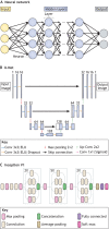
Fig. 1.
Fig. 1.
Neural networks and convolutional neural networks for bioimage analysis. (A) Schematic of a typical NN composed of an input layer (green), hidden layers (blue) and an output layer (red). Each layer is composed of neurons connected to each other. (B) Schematic of a U-net architecture as used in McGinn et al. (2021) for the segmentation of cells and nuclei in mouse epithelial tissues. U-net is amongst the most popular and efficient CNN models used for bioimage analysis and is designed using ‘convolutional’, ‘pooling’ and ‘dense’ layers as key building blocks (see Glossary, Box 1). U-net follows a symmetric encoder-decoder architecture resulting in a characteristic U-shape. Along the encoder path, the first branch of the U, the input image is progressively compacted, leading to a representation with reduced spatial information but increased feature information. Along the decoder path, the second branch of the U, feature and spatial information are combined with information from the encoder path, enforcing the model to learn image characteristics at various spatial scales. (C) Schematic of an Inception V1 architecture, also called ‘GoogleLeNet’. Inception V1 is a typical CNN architecture for image classification tasks. For example, it has been used to classify early human embryos images with very high accuracy (Khosravi et al., 2019). It is designed around a repetitive architecture made of so-called ‘inception blocks’, which apply several ‘convolutional’ and max ‘pooling’ layers (see Glossary, Box 1) to their input before concatenating together all generated feature maps.
Fig. 2.
Fig. 2.
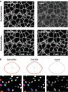
Deep learning methods applied to developmental biology applications. (A) A simulated ground truth cell membrane image is artificially degraded with noise. Denoised outputs obtained using Noise2Noise and Noise2Void are shown at the bottom, along with their average peak signal-to-noise values (PSNR; higher values translate to sharper, less-noisy images). Image adapted from Krull et al. (2019). (B) Fluorescence microscopy cell nuclei image from the Kaggle 2018 Data Science Bowl (dataset: BBBC038v1; Caicedo et al., 2019) segmented with StarDist (Schmidt et al., 2018), in which objects are represented as star-convex polygons, and with SplineDist, in which objects are described as a planar spline curve. Image adapted from Mandal and Uhlmann (2021).

Similar articles

Cited by

References

    1. Adebayo, J., Gilmer, J., Muelly, M., Goodfellow, I., Hardt, M. and Kim, B. (2018). Sanity checks for saliency maps. In Proceedings of NeurIPS 2018.
    1. Arganda-Carreras, I., Kaynig, V., Rueden, C., Eliceiri, K. W., Schindelin, J., Cardona, A. and Sebastian Seung, H. (2017). Trainable Weka Segmentation: a machine learning tool for microscopy pixel classification. Bioinformatics 33, 2424-2426. 10.1093/bioinformatics/btx180 - DOI - PubMed
    1. Argelaguet, R., Cuomo, A. S. E., Stegle, O. and Marioni, J. C. (2021). Computational principles and challenges in single-cell data integration. Nat. Biotechnol. [Epub ahead of print]. 10.1038/s41587-021-00895-7 - DOI - PubMed
    1. Ash, J. T., Darnell, G., Munro, D. and Engelhardt, B. E. (2021). Joint analysis of expression levels and histological images identifies genes associated with tissue morphology. Nat. Commun. 12, 1609. 10.1038/s41467-021-21727-x - DOI - PMC - PubMed
    1. Barber, R. F. and Candès, E. J. (2019). A knockoff filter for high-dimensional selective inference. Annals of Statistics 47, 2504-2537. 10.1214/18-AOS1755 - DOI

Publication types