

Bullous Scabies: Clinical, Dermoscopic, and Pathologic Characteristics of Ten Patients
- PMID: 34491217
- PMCID: PMC8641337
- DOI: 10.4269/ajtmh.21-0516
Bullous Scabies: Clinical, Dermoscopic, and Pathologic Characteristics of Ten Patients
Abstract
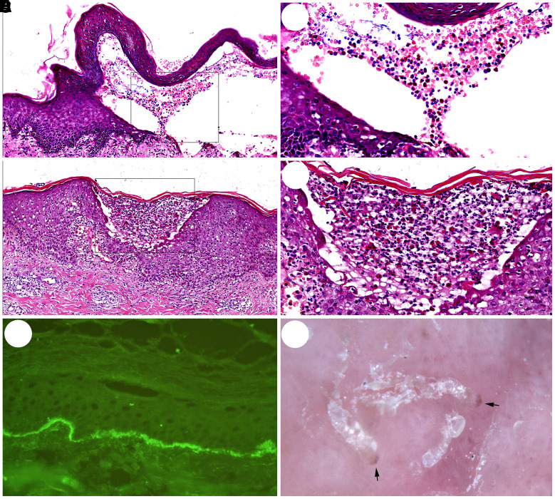
Bullous scabies (BS) is a rare atypical clinical variant of scabies and is easily confused with bullous disorders. The diagnosis of BS is always a challenge, and physicians often misdiagnose BS patients. Patients with BS admitted from 2012 to 2020 were enrolled in this study. The clinical, dermoscopic, and pathological characteristics of the patients were analyzed retrospectively. Ten patients with BS were enrolled in this study. Seven of the 10 patients were male. The bullae were most commonly found on the thighs and arms (80% of patients). Only 30% of patients (3/10) tested positive for mites and/or eggs by the initial skin scraping, but 100% (5/5) of the patients who received dermoscopy tested positive. Among these 10 patients, only five received a skin biopsy. Subepidermal (4/5) and intraepidermal (1/5) bullae with eosinophil and neutrophil infiltration were observed in five patients. Direct immunofluorescence (DIF) indicated linear deposition of IgG in the basement membrane zone in three patients. Physicians should consider the possibility of BS in patients with blisters, pruritus, and poor response to corticosteroids. Dermoscopy should be prioritized for the differential diagnosis of BS to exclude other bullous disorders. Finally, a biopsy should be performed on each patient with bullae.
Figures

References
-
- Salavastru CM, Chosidow O, Boffa MJ, Janier M, Tiplica GS, 2017. European guideline for the management of scabies. J Eur Acad Dermatol Venereol 31: 1248–1253. - PubMed
MeSH terms
Substances
LinkOut - more resources
Full Text Sources
Medical
