

Long Noncoding RNA MIAT Modulates the Extracellular Matrix Deposition in Leiomyomas by Sponging MiR-29 Family
- PMID: 34491311
- PMCID: PMC8459448
- DOI: 10.1210/endocr/bqab186
Long Noncoding RNA MIAT Modulates the Extracellular Matrix Deposition in Leiomyomas by Sponging MiR-29 Family
Abstract
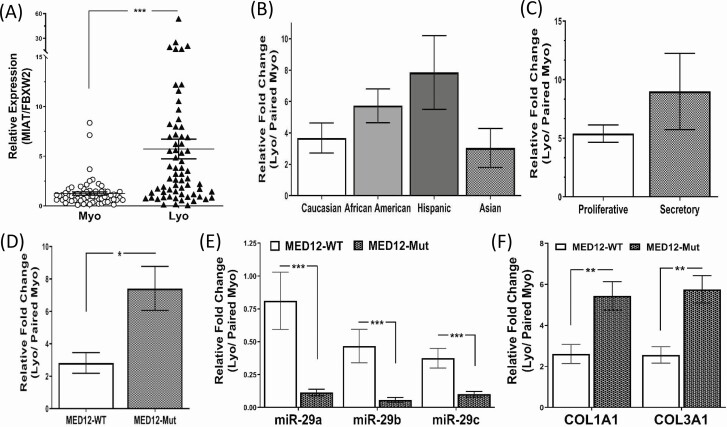
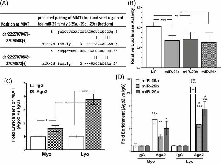
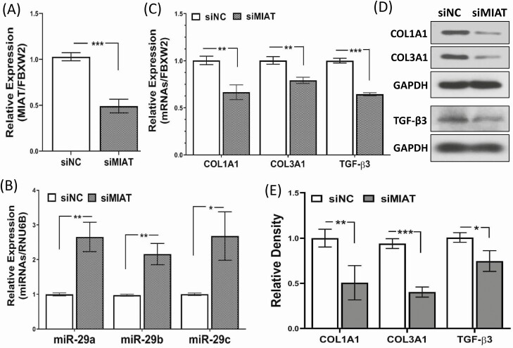
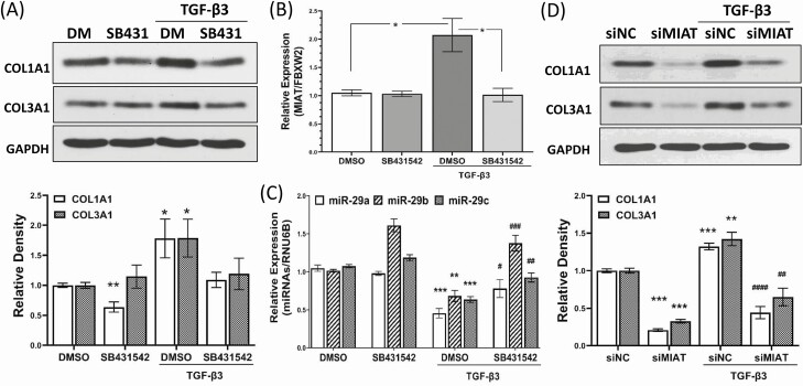
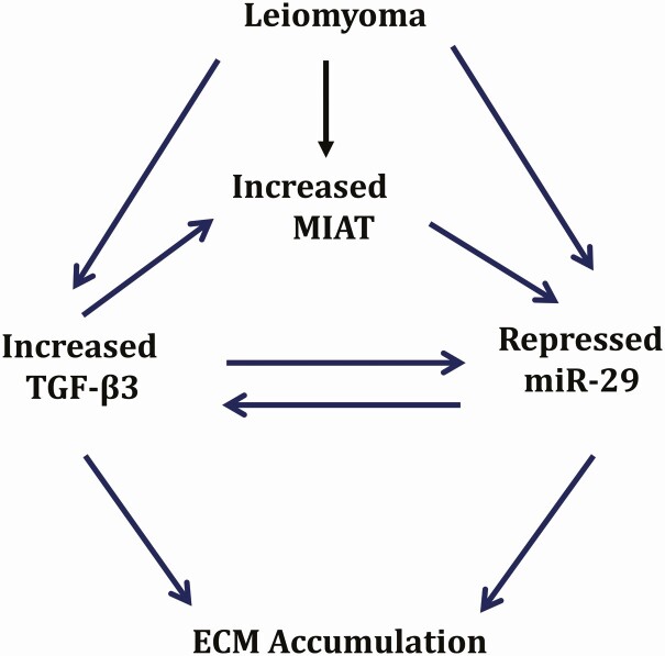
The objective of this study was to determine the expression and functional role of a long noncoding RNA (lncRNA) MIAT (myocardial infarction-associated transcript) in leiomyoma pathogenesis. Leiomyoma compared with myometrium (n = 66) expressed significantly more MIAT that was independent of race/ethnicity and menstrual cycle phase but dependent on MED12 (mediator complex subunit 12) mutation status. Leiomyomas bearing the MED12 mutation expressed higher levels of MIAT and lower levels of microRNA 29 family (miR-29a, -b, and -c) compared with MED12 wild-type leiomyomas. Using luciferase reporter activity and RNA immunoprecipitation analysis, MIAT was shown to sponge the miR-29 family. In a 3-dimensional spheroid culture system, transient transfection of MIAT siRNA in leiomyoma smooth muscle cell (LSMC) spheroids resulted in upregulation of miR-29 family and downregulation of miR-29 targets, collagen type I (COL1A1), collagen type III (COL3A1), and TGF-β3 (transforming growth factor β-3). Treatment of LSMC spheroids with TGF-β3 induced COL1A1, COL3A1, and MIAT levels, but repressed miR-29 family expression. Knockdown of MIAT in LSMC spheroids blocked the effects of TGF-β3 on the induction of COL1A1 and COL3A1 expression. Collectively, these results underscore the physiological significance of MIAT in extracellular matrix accumulation in leiomyoma.
Keywords: MED12 mutation; MIAT; TGF-β3; leiomyoma; miR-29.
© The Author(s) 2021. Published by Oxford University Press on behalf of the Endocrine Society. All rights reserved. For permissions, please e-mail: journals.permissions@oup.com.
Figures
Similar articles
-
Functional role of the long noncoding RNA X-inactive specific transcript in leiomyoma pathogenesis.Fertil Steril. 2021 Jan;115(1):238-247. doi: 10.1016/j.fertnstert.2020.07.024. Epub 2020 Oct 15. Fertil Steril. 2021. PMID: 33070965 Free PMC article.
-
Targeting the long non-coding RNA MIAT for the treatment of fibroids in an animal model.Clin Sci (Lond). 2024 Jun 19;138(12):699-709. doi: 10.1042/CS20240190. Clin Sci (Lond). 2024. PMID: 38817011 Free PMC article.
-
Mechanisms underlying aberrant expression of miR-29c in uterine leiomyoma.Fertil Steril. 2016 Jan;105(1):236-45.e1. doi: 10.1016/j.fertnstert.2015.09.020. Epub 2015 Oct 9. Fertil Steril. 2016. PMID: 26453978
-
MED12 and uterine smooth muscle oncogenesis: State of the art and perspectives.Eur J Cancer. 2015 Aug;51(12):1603-10. doi: 10.1016/j.ejca.2015.04.023. Epub 2015 May 30. Eur J Cancer. 2015. PMID: 26037152 Review.
-
The expression and potential regulatory function of microRNAs in the pathogenesis of leiomyoma.Semin Reprod Med. 2008 Nov;26(6):500-14. doi: 10.1055/s-0028-1096130. Epub 2008 Oct 24. Semin Reprod Med. 2008. PMID: 18951332 Free PMC article. Review.
Cited by
-
Differential Expression of MED12-Associated Coding RNA Transcripts in Uterine Leiomyomas.Int J Mol Sci. 2023 Feb 13;24(4):3742. doi: 10.3390/ijms24043742. Int J Mol Sci. 2023. PMID: 36835153 Free PMC article.
-
The Functional Role of the Long Non-Coding RNA LINCMD1 in Leiomyoma Pathogenesis.Int J Mol Sci. 2024 Oct 27;25(21):11539. doi: 10.3390/ijms252111539. Int J Mol Sci. 2024. PMID: 39519092 Free PMC article.
-
The in vivo effects of knockdown of long non-coding RNA XIST on fibroid growth and gene expression.FASEB J. 2024 Nov 15;38(21):e70140. doi: 10.1096/fj.202401982R. FASEB J. 2024. PMID: 39475327 Free PMC article.
-
In Vivo Effects of Bay 11-7082 on Fibroid Growth and Gene Expression: A Preclinical Study.Cells. 2024 Jun 24;13(13):1091. doi: 10.3390/cells13131091. Cells. 2024. PMID: 38994944 Free PMC article.
-
Potential prognostic value of a eight ferroptosis-related lncRNAs model and the correlative immune activity in oral squamous cell carcinoma.BMC Genom Data. 2022 Nov 16;23(1):80. doi: 10.1186/s12863-022-01097-z. BMC Genom Data. 2022. PMID: 36384476 Free PMC article.
References
-
- Doherty L, Mutlu L, Sinclair D, Taylor H. Uterine fibroids: clinical manifestations and contemporary management. Reprod Sci. 2014;21(9):1067-1092. - PubMed
-
- Bulun SE. Uterine fibroids. N Engl J Med. 2013;369(14):1344-1355. - PubMed
-
- Islam MS, Ciavattini A, Petraglia F, Castellucci M, Ciarmela P. Extracellular matrix in uterine leiomyoma pathogenesis: a potential target for future therapeutics. Hum Reprod Update. 2018;24(1):59-85. - PubMed
Publication types
MeSH terms
Substances
Grants and funding
LinkOut - more resources
Full Text Sources
Medical
Miscellaneous
