. 2021 Nov 16;89(12):e0037121.
doi: 10.1128/IAI.00371-21. Epub 2021 Sep 7.

Nitric Oxide Inhibition of Rickettsia rickettsii

Affiliations

Nitric Oxide Inhibition of Rickettsia rickettsii

Liam F Fitzsimmons et al. Infect Immun. .

Abstract

Rickettsia rickettsii, the causative agent of Rocky Mountain spotted fever, is an enzootic, obligate, intracellular bacterial pathogen. Nitric oxide (NO) synthesized by the inducible NO synthase (iNOS) is a potent antimicrobial component of innate immunity and has been implicated in the control of virulent Rickettsia spp. in diverse cell types. In this study, we examined the antibacterial role of NO on R. rickettsii. Our results indicate that NO challenge dramatically reduces R. rickettsii adhesion through the disruption of bacterial energetics. Additionally, NO-treated R. rickettsii cells were unable to synthesize protein or replicate in permissive cells. Activated, NO-producing macrophages restricted R. rickettsii infections, but inhibition of iNOS ablated the inhibition of bacterial growth. These data indicate that NO is a potent antirickettsial effector of innate immunity that targets energy generation in these pathogenic bacteria to prevent growth and subversion of infected host cells.

Keywords: Rickettsia; host defense; innate immunity; nitric oxide.


Figures
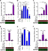
FIG 1
FIG 1
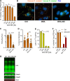
NO production is essential for clearance of R. rickettsii in activated J774 macrophages. (A) Culture supernatants were collected from J774 cells stimulated with LPS (1.5 ng/ml) or IFN-γ (15 ng/ml) for 24 h. The Griess reaction was used to determine nitrite concentrations (mean ± standard deviation [SD], n = 10). Additionally, monolayers were collected and analyzed by Western blotting for iNOS and GAPDH (representative images, n = 4). (B) Infectivity of R. rickettsii populations (PFU per milliliter) in infected J774 cells (MOI of 1 to 2) after 2 h (input) or 24 h with or without LPS or IFN-γ stimulation (mean ± SD, n = 6). (C) The Griess reaction was used to determine nitrite concentrations in culture supernatants from panel B, and Western blotting was used to examine iNOS and GAPDH expression (mean ± SD and representative images, n = 6). (D) J774 cells were stimulated as in panel A but with the addition of the iNOS inhibitor l-NIL (500 μM) and were analyzed similarly with the Griess reaction and Western blotting (mean ± SD and representative images, n = 7). (E) J774 cells were infected and stimulated as described for panel B but with l-NIL, and R. rickettsii populations were determined similarly (mean ± SD, n = 8). (F) Nitrite concentrations in culture supernatants and iNOS and GAPDH expression from samples described for panel E (mean ± SD and representative images, n = 8). Significance was determined with one-way ANOVA. ****, P < 0.0001.
FIG 2
FIG 2
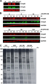
NO decreases R. rickettsii adhesion to host cells. (A) R. rickettsii was challenged with DEA (800 μM) or DEA-NO in BHI medium for 10 min at 34°C in 5% CO2, and infectivity was determined by plaque counts (mean ± SD, n = 4). (B) Immunofluorescence microscopy of external and internal R. rickettsii cells treated with DEA or DEA-NO (images are representative of three or four independent experiments). (C and D) Quantification of images in panel B. The total number of R. rickettsii bacteria per cell was determined (C) and the proportion of internalized R. rickettsii was calculated (D) (five images were quantified from three or four independent experiments). (E) Nucleotides were extracted from R. rickettsii cells that had been challenged with DEA or DEA-NO for 10 min in BHI medium. Extracts were neutralized, and ATP content was determined with firefly luciferase and luminescence measurements (mean ± SD, n = 3). (F). R. rickettsii cells were challenged with or without 800 μM DEA-NO for 10 min in BHI medium. Samples were diluted into BHI medium or BHI medium supplemented with 1 mM ATP and incubated at 34°C in 5% CO2 for 30 min, and plaque assays determined infectivity (mean ± SD, n = 4). (G) R. rickettsii cells were challenged with DEA or DEA-NO (1 mM in BHI medium) for 10 min at 34°C and were analyzed by Western blotting. Antibodies against spotted fever rickettsial LPS (LPS), rOmpA, rOmpB, and Sca1 were used to detect R. rickettsii outer membrane antigens (representative blots, n = 2). Statistical analyses were performed using one-way ANOVA. *, P < 0.05; ***, P < 0.001; ****, P <0.0001; ns, not significant.
FIG 3
FIG 3
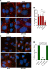
NO inhibits R. rickettsii intracellular growth. (A) Vero cells were infected for 24 h with R. rickettsii and then challenged with SPR or SPR-NO for 3 h. Select samples were analyzed for PFU or were given fresh medium for 21 h before being analyzed for PFU (mean ± SD, n = 3 or 6). (B) Vero cells were infected with R. rickettsii for 2 h, input samples were harvested, and the remaining samples were challenged with SPR or SPR-NO (150 mM) for 24 h prior to harvesting for PFU determinations (mean ± SD, n = 4). (C) Samples were treated as described in panel B but were analyzed by Western blotting with anti-rOmpA, anti-rOmpB, and anti-GAPDH antibodies. Relative levels of rOmpA and rOmpB were quantified and normalized to GAPDH loading controls (mean ± SD, n = 3; representative images are Fig. 4A). (D) Vero cells were infected with R. rickettsii for 2 h and then challenged with increasing amounts of SPR-NO. Samples were harvested at 0, 8, 16, and 24 hpt, analyzed by Western blotting, and quantified as described for panel C (mean ± SD, n = 5; representative images are Fig. 4B). Statistical analyses were performed using one- or two-way ANOVA. **, P < 0.01; ***, P < 0.001; ****, P  <0.0001; ns, not significant.
FIG 4
FIG 4
NO inhibits protein synthesis of intracellular R. rickettsii. (A) Vero cells were infected with R. rickettsii for 2 h, and select samples were collected immediately (IN) or were treated with 150 μM SPR or SPR-NO and then collected 24 h later. Samples were processed for Western blotting with anti-rOmpA, anti-rOmpB, and anti-GAPDH antibodies (representative blot, n = 3). (B) Vero cells were infected with R. rickettsii for 2 h and treated with increasing concentrations of SPR-NO. After 0, 8, 16, and 24 h of treatment, samples were collected for Western blotting. Anti-rOmpA, anti-rOmpB, and anti-GAPDH antibodies were used (representative images, n = 5). (C) Vero cells were infected with R. rickettsii for 24 h, treated with SPR or SPR-NO for 3 h, and then labeled with [35S]Met for 3 h. Samples were scraped into Laemmli sample buffer, boiled, and separated by SDS-PAGE, gels were dried and exposed to film, and autoradiograms were visualized (representative autoradiogram, n = 3).
FIG 5
FIG 5
NO disrupts host cell subversion by intracellular R. rickettsii. (A) Vero cells on coverslips were infected for 2 h with R. rickettsii and were harvested (Input) or were untreated (Untr) or treated with 150 μM SPR or SPR-NO for 12 h. Samples were visualized by immunofluorescence microscopy. rOmpB antibody 13-2, phalloidin-647, and DAPI, were used to detect R. rickettsii (green), F-actin (red), and DNA (blue), respectively (representative images, n = 3). (B) The proportions of R. rickettsii cells with evident actin tails were calculated from data in panel A (mean ± SD, n = 6). (C) Vero cells were treated as described for panel B, but samples were inactivated at 17 hpt, and rOmpB antibody 13-2, anti-TGN46, and DAPI were used to detect R. rickettsii (green), the TGN (red/orange), and DNA (blue), respectively (representative images, n = 3). (D) Cells were assessed for intact or dispersed TGN in images from C, and the proportions of cells with intact TGN were calculated (mean ± SD, n = 3). Statistical analyses were performed using one-way ANOVA. ****, P  <0.0001; ns, not significant.

References

    1. Paddock CD, Denison AM, Lash RR, Liu L, Bollweg BC, Dahlgren FS, Kanamura CT, Angerami RN, Pereira dos Santos FC, Brasil Martines R, Karpathy SE. 2014. Phylogeograpy of Rickettsia rickettsii genotypes associated with fatal Rocky Mountain spotted fever. Am J Trop Med Hyg 91:589–597. 10.4269/ajtmh.14-0146. - DOI - PMC - PubMed
    1. Bogdan C. 2015. Nitric oxide synthase in innate and adaptive immunity: an update. Trends Immunol 36:161–178. 10.1016/j.it.2015.01.003. - DOI - PubMed
    1. Bhattacharya ST, Bayakly N, Lloyd R, Benson MT, Davenport J, Fitzgerald ME, Rothschild M, Lamoreaux WJ, Coons LB. 2000. Nitric oxide synthase and cGMP activity in the salivary glands of the American dog tick Dermacentor variabilis. Exp Parasitol 94:111–120. 10.1006/expr.1999.4477. - DOI - PubMed
    1. Porrini C, Ramarao N, Tran SL. 2020. Dr. NO and Mr. Toxic: the versatile role of nitric oxide. Biol Chem 401:547–572. 10.1515/hsz-2019-0368. - DOI - PubMed
    1. Cinelli MA, Do HT, Miley GP, Silverman RB. 2020. Inducible nitric oxide synthase: regulation, structure, and inhibition. Med Res Rev 40:158–189. 10.1002/med.21599. - DOI - PMC - PubMed

Publication types

MeSH terms

LinkOut - more resources