

Metabolic Plasticity Aids Amphotropism of Coxiella burnetii
- PMID: 34491791
- PMCID: PMC8594591
- DOI: 10.1128/IAI.00135-21
Metabolic Plasticity Aids Amphotropism of Coxiella burnetii
Abstract
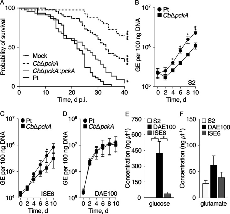
Coxiella burnetii, the causative agent of query (Q) fever in humans, is an obligate intracellular bacterium. C. burnetii can naturally infect a broad range of host organisms (e.g., mammals and arthropods) and cell types. This amphotropic nature of C. burnetii, in combination with its ability to utilize both glycolytic and gluconeogenic carbon sources, suggests that the pathogen relies on metabolic plasticity to replicate in nutritionally diverse intracellular environments. To test the significance of metabolic plasticity in C. burnetii host cell colonization, C. burnetii intracellular replication in seven distinct cell lines was compared between a metabolically competent parental strain and a mutant, CbΔpckA, unable to undergo gluconeogenesis. Both the parental strain and CbΔpckA mutant exhibited host cell-dependent infection phenotypes, which were influenced by alterations to host glycolytic or gluconeogenic substrate availability. Because the nutritional environment directly impacts host cell physiology, our analysis was extended to investigate the response of C. burnetii replication in mammalian host cells cultivated in a novel physiological medium based on the nutrient composition of mammalian interstitial fluid, interstitial fluid-modeled medium (IFmM). An infection model based on IFmM resulted in exacerbation of a replication defect exhibited by the CbΔpckA mutant in specific cell lines. The CbΔpckA mutant was also attenuated during infection of an animal host. Overall, the study underscores that gluconeogenic capacity aids C. burnetii amphotropism and that the amphotropic nature of C. burnetii should be considered when resolving virulence mechanisms in this pathogen.
Keywords: Coxiella burnetii; amphotropism; gluconeogenesis; glycolysis; interstitial fluid; intracellular parasites; metabolic plasticity; obligate; physiological medium; virulence.
Figures
Similar articles
-
Physicochemical and Nutritional Requirements for Axenic Replication Suggest Physiological Basis for Coxiella burnetii Niche Restriction.Front Cell Infect Microbiol. 2017 May 31;7:190. doi: 10.3389/fcimb.2017.00190. eCollection 2017. Front Cell Infect Microbiol. 2017. PMID: 28620582 Free PMC article.
-
EirA Is a Novel Protein Essential for Intracellular Replication of Coxiella burnetii.Infect Immun. 2020 May 20;88(6):e00913-19. doi: 10.1128/IAI.00913-19. Print 2020 May 20. Infect Immun. 2020. PMID: 32205404 Free PMC article.
-
Critical Role for Molecular Iron in Coxiella burnetii Replication and Viability.mSphere. 2020 Jul 22;5(4):e00458-20. doi: 10.1128/mSphere.00458-20. mSphere. 2020. PMID: 32699121 Free PMC article.
-
Life on the outside: the rescue of Coxiella burnetii from its host cell.Annu Rev Microbiol. 2011;65:111-28. doi: 10.1146/annurev-micro-090110-102927. Annu Rev Microbiol. 2011. PMID: 21639786 Review.
-
From cell culture to cynomolgus macaque: infection models show lineage-specific virulence potential of Coxiella burnetii.J Med Microbiol. 2019 Oct;68(10):1419-1430. doi: 10.1099/jmm.0.001064. J Med Microbiol. 2019. PMID: 31424378 Review.
Cited by
-
Primary Murine Macrophages as a Tool for Virulence Factor Discovery in Coxiella burnetii.Microbiol Spectr. 2022 Aug 31;10(4):e0248421. doi: 10.1128/spectrum.02484-21. Epub 2022 Aug 1. Microbiol Spectr. 2022. PMID: 35913176 Free PMC article.
-
Recent advances in genetic systems in obligate intracellular human-pathogenic bacteria.Front Cell Infect Microbiol. 2023 Jun 19;13:1202245. doi: 10.3389/fcimb.2023.1202245. eCollection 2023. Front Cell Infect Microbiol. 2023. PMID: 37404720 Free PMC article. Review.
-
Coxiella burnetii protein CBU2016 supports CCV expansion.Pathog Dis. 2024 Feb 7;82:ftae018. doi: 10.1093/femspd/ftae018. Pathog Dis. 2024. PMID: 39138067 Free PMC article.
-
Metabolism and physiology of pathogenic bacterial obligate intracellular parasites.Front Cell Infect Microbiol. 2024 Mar 22;14:1284701. doi: 10.3389/fcimb.2024.1284701. eCollection 2024. Front Cell Infect Microbiol. 2024. PMID: 38585652 Free PMC article. Review.
-
The endogenous Coxiella burnetii plasmid encodes a functional toxin-antitoxin system.Mol Microbiol. 2022 Dec;118(6):744-764. doi: 10.1111/mmi.15001. Epub 2022 Nov 28. Mol Microbiol. 2022. PMID: 36385554 Free PMC article.
References
-
- Anderson A, Bijlmer H, Fournier P-E, Graves S, Hartzell J, Kersh GJ, Limonard G, Marrie TJ, Massung RF, McQuiston JH, Nicholson WL, Paddock CD, Sexton DJ. 2013. Diagnosis and management of Q fever–United States, 2013: recommendations from CDC and the Q Fever Working Group. MMWR Recomm Rep 62:1–30. - PubMed
-
- Melenotte C, Protopopescu C, Million M, Edouard S, Carrieri MP, Eldin C, Angelakis E, Djossou F, Bardin N, Fournier P-E, Mège J-L, Raoult D. 2018. Clinical features and complications of Coxiella burnetii infections from the French National Reference Center for Q Fever. JAMA Netw Open 1:e181580. 10.1001/jamanetworkopen.2018.1580. - DOI - PMC - PubMed
-
- Delsing CE, Kullberg BJ, Bleeker-Rovers CP. 2010. Q fever in the Netherlands from 2007 to 2010. Neth J Med 68:382–387. - PubMed
Publication types
MeSH terms
Substances
Grants and funding
LinkOut - more resources
Full Text Sources
