Review
. 2021 Dec;94(1128):20210099.
doi: 10.1259/bjr.20210099. Epub 2021 Sep 7.

Mimics of perineural tumor spread in the head and neck

Affiliations
Review

Mimics of perineural tumor spread in the head and neck

Manoj Tanwar et al. Br J Radiol. 2021 Dec.

Abstract

Perineural spread (PNS) is an important potential complication of head and neck malignancy, as it is associated with decreased survival and a higher risk of local recurrence and metastasis. There are many review articles focused on the imaging findings of PNS. However, a false-positive diagnosis of PNS can be just as harmful to the patient as an overlooked case. In this manuscript, we delineate and classify various imaging mimics of PNS. Mimics can be divided into the following categories: normal variants (including vascular structures and failed fat suppression), infections, inflammatory disease (including granulomatous disease and demyelination), neoplasms, and post-traumatic/surgical changes. Knowledge of potential mimics of PNS will prevent false-positive imaging interpretation, and enable appropriate oncologic management.


Figures

Figure 1.
Figure 1.
Adenoid cystic carcinoma of the palate: axial T1 +C images show tumor extending superiorly to involve multiple cranial nerves. (a) Irregular thickening and enhancement of the right cavernous sinus is a hallmark of perineural tumor spread. The maxillary division of CN V within the foramen rotundum is enlarged (long arrow), and abnormal enhancement extends into the infraorbital nerve (arrowhead). (b) On a slightly inferior image, abnormal infiltrative enhancement is seen within the pterygopalatine fossa (short arrow) and foramen lacerum (arrowhead).
Figure 2.
Figure 2.
Nasopharyngeal squamous cell carcinoma: coronal T1 +C images show (a) avidly enhancing tumor within foramen ovale (long arrow). (b) There is also tumor extension superiorly into the Meckel’s cave (short arrow).
Figure 3.
Figure 3.
Normal prominent pterygoid venous plexus: axial CECT demonstrates strikingly asymmetric plexus of veins surrounding the left medial and lateral pterygoid muscles (arrow), which can be easily mistaken for a tumor. CECT, contrast-enhanced CT.
Figure 4.
Figure 4.
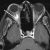
Failed fat suppression: axial T1 +C image acquired with fat saturation depicts a tumefactive area of enhancement surrounding the optic nerve (arrow). This artifact resulting from the failure of fat saturation due to field distortion from dental amalgam can be easily mistaken for an abnormal lesion.
Figure 5.
Figure 5.
Bell’s palsy: axial T1 +C image shows abnormal enhancement in the labyrinthine segment and anterior genu of the right facial nerve (arrow). Although PNS can have skip lesions, enhancement exclusive to the internal auditory canal is unlikely to represent PNS. PNS, perineural spread.
Figure 6.
Figure 6.
Ramsay Hunt syndrome: herpetic infection of VII and VIII CNs in a patient who presented with complaints of vertigo. Linear enhancement within the right internal auditory canal (arrow) as seen on the coronal T1 +C image will favor viral infection. A nodular or fusiform enhancement could raise suspicion for tumor spread.
Figure 7.
Figure 7.
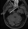
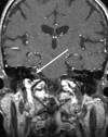
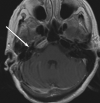
Trauma with superimposed bacterial orbital cellulitis: infection tracking along intracranial nerves can appear similar to perineural tumor spread. In this patient, bacterial cellulitis had tracked posteriorly through the superior orbital fissure (arrowhead) and foramen rotundum along with CN V (long arrow) to its root entry zone on the axial (a) and coronal (b) T1 +C images. This resulted in a rim-enhancing bacterial abscess (short arrow) in the left brachium pontis and cerebellum. A tumor spreading via the ophthalmic division of CN V could behave in a similar fashion.
Figure 8.
Figure 8.
Invasive fungal sinusitis secondary to mucormycosis: axial T1 +C images show (a) diffuse enhancement near the sphenopalatine foramen (long arrow), vidian canal (short arrow), and foramen ovale (arrowhead). (b) More superiorly, the infection involves foramen rotundum (arrow). This appearance is remarkably similar to the cases with perineural tumor spread.
Figure 9.
Figure 9.
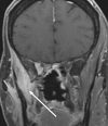
Invasive fungal sinusitis due to mucormycosis: axial (a) and coronal (b) CECT images show left maxillary sinus fungal disease with associated enhancement in the sphenopalatine foramen (arrowhead), pterygopalatine fossa (short arrow), and foramen rotundum (long arrow). PNS can result in a similar pattern of enhancement.
Figure 10.
Figure 10.
Granulomatosis with polyangiitis: axial (a) and coronal (b) T1 +C images demonstrate abnormal enhancement in the right orbit (arrow), orbital apex (arrowhead), and downward extension into the infratemporal fossa (short arrow). An orbital tumor with associated involvement of trigeminal nerve divisions can result in a similar pattern of enhancement.
Figure 11.
Figure 11.
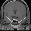
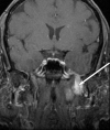
Neurosarcoidosis: involvement of the skull base and adjacent meninges by sarcoid may be reminiscent of perineural tumor spread. On axial (a) and coronal (b) T1 +C images, inflammatory changes extend through and expand left foramen rotundum (arrowhead) as well as foramen ovale (short arrow). There is also asymmetric enlargement of the cavernous sinus (long arrow). In a different patient, these findings would be typical for the perineural spread of tumor.
Figure 12.
Figure 12.
CIDP: coronal T1 +C image shows enhancement in the V3 segments of both trigeminal nerves (arrows) which can be confused with PNS. However, bilateral involvement would be uncommon in tumor spread. CIDP, chronic inflammatory demyelinating polyneuropathy; PNS, perineural spread.
Figure 13.
Figure 13.
Jugular canal schwannomas: that cross the skull base can expand the traversing foramina, such as the foramen ovale in this case on the coronal T1 +C image (arrow). This mass demonstrates a more lobular appearance than expected for perineural spread.
Figure 14.
Figure 14.
Paragangliomas: usually arise in predictable locations, but occasionally they may arise in regions typically associated with perineural tumor spread, such as in the vicinity of stylomastoid foramen on this axial T1 +C image (arrow).
Figure 15.
Figure 15.
Plexiform neurofibroma in neurofibromatosis type I. (a) Axial T1 +C image shows an elongated heterogeneously enhancing lesion (arrow) within the lateral aspect of the right orbit extending through the superior orbital fissure. This would be the expected course of perineural tumor spread along with CN III. (b) Axial T2 weighted image further confirms the presence of an elongated hyperintense lesion (arrow).
Figure 16.
Figure 16.
Esophageal carcinoma with intracranial leptomeningeal metastases: T1 +C images at two levels demonstrate abnormal enhancement along multiple nerves, such as III (long arrow, (a) and VII/ VIII nerve complex (short arrow, b). These linear enhancements can be easily confused with PNS, however multifocal involvement favors the leptomeningeal disease. PNS, perineural spread.
Figure 17.
Figure 17.
Meningioma: axial (a) and coronal (b) T1 +C images show a homogenously enhancing mass within the left cavernous sinus (arrows). The mass encases the cavernous internal carotid artery, but the flow void remains preserved. Perineural spread may have a similar imaging appearance with cavernous sinus expansion, and bowing of lateral sinus wall.
Figure 18.
Figure 18.
Acute denervation muscle enhancement: coronal T1 +C image shows abnormal enhancement in the right muscles of mastication (arrow) secondary to acute denervation injury. This enhancement is however not a tumor, and will only be seen in the acute stage of denervation. Perineural tumor spread via branches of the mandibular division of CN V could give rise to a similar appearance.
Figure 19.
Figure 19.
Post-surgical changes: this patient had undergone a translabrynthine approach right vestibular schwannoma resection. Axial T1 +C image demonstrates asymmetric enhancement along the expected tympanic segment of the right facial nerve (arrow), which can be easily mistaken for perineural tumor spread. This thin linear enhancement will be expected to remain stable, or decrease over follow-up studies.

Similar articles

Cited by

References

    1. Wild C. P, Weiderpass E, Stewart B. W. eds. World cancer report: cancer research for cancer prevention. Lyon, France: International Agency for Research on Cancer; 2020. .
    1. Huang SH, O'Sullivan B. Overview of the 8th edition TNM classification for head and neck cancer. Curr Treat Options Oncol 2017; 18: 40. doi: 10.1007/s11864-017-0484-y - DOI - PubMed
    1. Liebig C, Ayala G, Wilks JA, Berger DH, Albo D. Perineural invasion in cancer: a review of the literature. Cancer 2009; 115: 3379–91. doi: 10.1002/cncr.24396 - DOI - PubMed
    1. Bakst RL, Glastonbury CM, Parvathaneni U, Katabi N, Hu KS, Yom SS. Perineural invasion and perineural tumor spread in head and neck cancer. Int J Radiat Oncol Biol Phys 2019; 103: 1109–24. doi: 10.1016/j.ijrobp.2018.12.009 - DOI - PubMed
    1. Borges A, Casselman J. Imaging the cranial nerves: Part I: methodology, infectious and inflammatory, traumatic and congenital lesions. Eur Radiol 2007; 17: 2112–25. doi: 10.1007/s00330-006-0575-9 - DOI - PubMed

MeSH terms