Review
. 2021 Nov 1;94(1127):20210281.
doi: 10.1259/bjr.20210281. Epub 2021 Sep 7.

Imaging of acute pelvic pain

Affiliations
Review

Imaging of acute pelvic pain

Marijana Basta Nikolic et al. Br J Radiol. .

Abstract

Acute pelvic pain (APP) requires urgent medical evaluation and treatment. Differential diagnosis of APP is broad, including a variety of gynecologic and non-gynecologic/ urinary, gastrointestinal, vascular and other entities. Close anatomical and physiological relations of pelvic structures, together with similar clinical presentation of different disorders and overlapping of symptoms, especially in the emergency background, make the proper diagnosis of APP challenging. Imaging plays a crucial role in the fast and precise diagnosis of APP. Ultrasonography is the first-line imaging modality, often accompanied by CT, while MRI is utilized in specific cases, using short, tailored protocols. Recognizing the cause of APP in females is a challenging task, due to the wide spectrum of possible origin and overlap of their imaging features. Therefore, the radiologist has to be familiar with the possible causes of APP, and, relying on clinical presentation, together with laboratory findings, choose the best imaging strategy in order to establish a fast and accurate diagnosis.


Figures

Figure 1.
Figure 1.
Diagnostic approach to the most common causes of APP -ultrasonography. APP, acute pelvic pain; CECT, contrast-enhanced computed tomography; DG, diagnosis; GI, Gastrointestinal; IUP, Intrauterine pregnancy; NECT, non-enhanced computed tomography.
Figure 2.
Figure 2.
Rupture of corpus luteum cyst. Contrast-enhanced CT (a, b) shows multiple cysts with thick, enhancing walls (arrowhead), corresponding to the “ring of fire“ sign on ultrasound, and active extravasation of contrast agent (arrow), accompanied by hematoperitoneum (asterisk). (c) Intraoperative specimen.
Figure 3.
Figure 3.
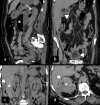
Pelvic inflammatory disease. Contrast-enhanced CT (a, b) shows slightly enlarged, inhomogenous left ovary (arrowhead), together with left parauterine tubular structure with enhancing walls (arrow), resembling inflamed fallopian tube, accompanied by ascites (asterisk). MRI (c, d) performed 3 days later confirmed oophoritis (arrowhead), however, parauterine mass was confirmed to be sigmoid colon loop (arrow).
Figure 4.
Figure 4.
Ovarian torsion. Inhomogenous retrouterine pelvic mass (arrowhead) presenting edematous, displaced ovary due to ovarian torsion, accompanied by ascites (arrow).
Figure 5.
Figure 5.
Degenerated myoma. Coronal (a), axial (b), and sagittal (c) contrast-enhanced CT shows large myoma (asterisk) with signs of extensive degeneration.
Figure 6.
Figure 6.
Torsion of pedunculated myoma. (a) Coronal contrast-enhanced CT of torsion of pedunculated subserous myoma (arrowhead). Differential diagnosis towards torsion of an ovarian lesion can be challenging. Intraoperative specimen (b) and T2W MRI (c, d) of partly necrotic solid ovarian tumor (asterisk), due to torsion, with typical ”whirlpool“ sign (arrow).
Figure 7.
Figure 7.
Patient in the second trimester of pregnancy presenting with acute abdominopelvic pain. Coronal (a, b) and sagittal (c) T2W MRI show partly degenerated intramural myoma (asterisk), placenta previa (arrowhead), and right hydronephrosis (arrow), caused by compression of the right ureter by the enlarged uterus. Acute pain in this patient is caused by hydronephrosis.
Figure 8.
Figure 8.
Ectopic pregnancy. (a, b) Ruptured right tubal ectopic pregnancy (arrowhead) with massive hematoperitoneum (asterisk). (c, d) Unperforated left tubal ectopic pregnancy (arrow)—CT shows gestational sac with no signs of rupture.
Figure 9.
Figure 9.
Ovarian hypertimulation syndrome. Ultrasonography depicts enlarged ovaries with multiple follicular cysts arranged in a spoke-wheel pattern.
Figure 10.
Figure 10.
Gynecological malignancies presenting as APP. (a, b and c) Cervical cancer. (a) Ultrasound of cervical cancer (asterisk) causing cervical stenosis and consequent pyometra (arrowhead). (b) CT shows a distended uterine cavum filled with hypodense thick fluid (arrowhead). (c) Intraoperative specimen confirms the diagnosis of pyometra caused by tumor inducing cervical stenosis. (d) and (e) hemorrhagic ovarian metastasis presenting as T1FS hyperintense ovarian mass (arrow). Pre- (d) and post-contrast (e) T1FS tomograms.
Figure 11.
Figure 11.
Calculus (arrowhead) in the distant segment of the right ureter, causing obstruction and proximal dilatation of ureter and hydronephrosis. Pelvic ureterolithiasis can easily be misdiagnosed as phleboliths (arrow).
Figure 12.
Figure 12.
Pyelonephritis. Contrast-enhanced CT appreciation of bilateral hypodense wedge-shaped areas within thickened renal parenchyma.
Figure 13.
Figure 13.
Non-contrast CT in evaluation of urinary emergencies. (a) Patient with emphysematous pyelonephritis presenting with gas collections in pyelon (asterisk) and upper calyx (arrow). (b) The same patient with gas collections in the wall of the gallbladder and urinary bladder (arrowheads)—simultaneous emphysematous cholecystitis and cystitis. (c, d) Perinephric abscess—thick perirenal collection (asterisk) with gas particles (arrowhead).
Figure 14.
Figure 14.
Appendicitis—a spectrum of appearances. (a) Enhancement of appendiceal wall (arrowhead) with periappendicular fat stranding and abscess collection (asterisk) below the insertion of appendix to the coecum. (b) Extraluminal appendicolith (arrowhead) inside periappendicular abscess. (c) Intraluminal appendicolith (arrowhead) at the insertion of the appendix—note the intraluminal air in the proximal appendix, extraluminal air bubble (arrow), contrast enhancement defect of the appendiceal wall just posterior to the air bubble, and intraperitoneal liquid (asterisk). (d) Appendicitis in a large ventral hernia—contrast enhancement of appendiceal wall (arrowhead), periappendicular fat stranding and intraperitoneal free fluid (arrow). (e) The same patient as in (d) 7 years before with horizontally positioned appendix with no signs of inflammation. (f) Imaging pitfalls—dense appendicoliths or contrast from previous imaging causing artifacts which make evaluation of the appendiceal wall and adjacent fat difficult with no evident signs of appendicitis (operative findings indicated acute gangrenous appendicitis).
Figure 15.
Figure 15.
Acute diverticulitis of sigmoid colon—a spectrum of appearances. (a) Non-complicated inflamed diverticulum of the sigmoid colon (arrowhead) with stranding of adjacent fat and edema of the sigmoid wall. (b) Two simultaneous diverticula with signs of non-complicated inflammation (arrowheads). (c) Liquid–fluid collection contained within mesosigmoid fat not bigger than 4 cm (arrowhead).
Figure 16.
Figure 16.
Epiploic appendagitis. Ovoid fatty formation with fat stranding as a sign of edema next to the sigmoid wall. Demarcation of a hyperattenuating ring of visceral peritoneum (arrowhead). Note the lack of edema of the sigmoid wall contrary to the findings of acute diverticulitis.
Figure 17.
Figure 17.
Necrosis of terminal ileum. (a, b) Submucous fat deposition and edema (arrow) of terminal ileum and colon with mucosal hyperemia (arrowhead); air collection in terminal ileum wall as a sign of necrosis (asterisk) and free air bubbles next to the ileal and cecal wall.
Figure 18.
Figure 18.
Rupture of the right common iliac artery fusiform aneurysm. (a) VRT MIP - active extravasation of contrast material (arrow). (b) Place of the rupture of the arterial wall (arrowhead) and massive retroperitoneal hematoma (asterisk).

References

    1. Stoker J, van Randen A, Laméris W, Boermeester MA, et al. . Imaging patients with acute abdominal pain. Radiology 2009; 253: 31–46. doi: 10.1148/radiol.2531090302 - DOI - PubMed
    1. Otoni JC, Noschang J, Okamoto TY, Vieira DR, Petry MSM, de Araujo Ramos L, et al. . Role of computed tomography at a cancer center emergency department. Emerg Radiol 2017; 24: 113–7. doi: 10.1007/s10140-016-1449-3 - DOI - PubMed
    1. Zafar N, Kupesic Plavsic S. Role of ultrasound in the evaluation of acute pelvic pain in nonpregnant reproductive age patients. Reprod Age Patients Donald Sch J Ultrasound Obs Gynecol 2012; 6: 207–17. doi: 10.5005/jp-journals-10009-1244 - DOI
    1. Alt C, Bharwani N, Brunesch L, Stanza FM, Ma D, El SRF. ESUR quick guide to female pelvis imaging. Lisboa, Portugal: ESUR; 2019.
    1. Scheirey CD, Fowler KJ, Therrien JA, Kim DH, Al-Refaie WB, Camacho MA, et al. . ACR Appropriateness Criteria® Acute Nonlocalized Abdominal Pain. J Am Coll Radiol 2018; 15: S217–31. doi: 10.1016/j.jacr.2018.09.010 - DOI - PubMed

MeSH terms