. 2021 Sep;102(9):001651.
doi: 10.1099/jgv.0.001651.

Murine norovirus virulence factor 1 (VF1) protein contributes to viral fitness during persistent infection

Affiliations

Murine norovirus virulence factor 1 (VF1) protein contributes to viral fitness during persistent infection

Constantina Borg et al. J Gen Virol. 2021 Sep.

Abstract

Murine norovirus (MNV) is widely used as a model for studying norovirus biology. While MNV isolates vary in their pathogenesis, infection of immunocompetent mice mostly results in persistent infection. The ability of a virus to establish a persistent infection is dependent on its ability to subvert or avoid the host immune response. Previously, we described the identification and characterization of virulence factor 1 (VF1) in MNV, and demonstrated its role as an innate immune antagonist. Here, we explore the role of VF1 during persistent MNV infection in an immunocompetent host. Using reverse genetics, we generated MNV-3 viruses carrying a single or a triple termination codon inserted in the VF1 ORF. VF1-deleted MNV-3 replicated to comparable levels to the wildtype virus in tissue culture. Comparative studies between MNV-3 and an acute MNV-1 strain show that MNV-3 VF1 exerts the same functions as MNV-1 VF1, but with reduced potency. C57BL/6 mice infected with VF1-deleted MNV-3 showed significantly reduced replication kinetics during the acute phase of the infection, but viral loads rapidly reached the levels seen in mice infected with wildtype virus after phenotypic restoration of VF1 expression. Infection with an MNV-3 mutant that had three termination codons inserted into VF1, in which reversion was suppressed, resulted in consistently lower replication throughout a 3 month persistent infection in mice, suggesting a role for VF1 in viral fitness in vivo. Our results indicate that VF1 expressed by a persistent strain of MNV also functions to antagonize the innate response to infection. We found that VF1 is not essential for viral persistence, but instead contributes to viral fitness in mice. These data fit with the hypothesis that noroviruses utilize multiple mechanisms to avoid and/or control the host response to infection and that VF1 is just one component of this.

Keywords: VF1; accessory protein; interferon response; norovirus; viral persistence.


Conflict of interest statement

The authors declare that there are no conflicts of interest.

Figures

Fig. 1.
Fig. 1.
In vitro characterization of MNV-3 VF1. (a) A schematic representation of the MNV genome, showing the four ORFs that encode the non-structural proteins NS1/2–7, VP1, VP2 and VF1. (b) Western blot analysis of VF1 expression following a 12 h infection with MNV-1 (1-WT) and MNV-3 (3-WT) in RAW264.7 cells (m.o.i.=5). Mock inoculated samples were treated with Dulbecco's modified Eagle medium (DMEM). (c) An alignment of VF1 protein sequences from MNV-1 (acute, lab-adapted strain), MNV-3 (persistent, lab-adapted strain), CR6 (persistent, lab-adapted strain) and WM6 (persistent, wild strain). VF1 encoded by MNV-3 and MNV-1 are 89 % identical. (d) Confocal microscopy of MEFs transfected with plasmids expressing enhanced green fluorescent protein (EGFP), or EGFP fused to the N or C terminus of MNV-3 VF1. Green indicates EGFP, red indicates MitoTracker and blue denotes DAPI. (e) Biochemical fractionation of BV-2 cells infected with MNV-1 (1-WT) and MNV-3 (3-WT) (m.o.i.=5). Mock inoculated cells were treated with DMEM. Lysates were collected 12 h post-infection and mitochondrial (mito.) and cytoplasmic (cyto.) fractions were enriched as described in the text.
Fig. 2.
Fig. 2.
MNV-3 VF1 is dispensable for replication in cell lines. (a, b) Schematic representations of the MNV subgenomic RNA indicating sites of stop codon placements for MNV-3 M1 and MNV-3 M3. Stop codon mutations are nonsense for VF1 as they introduce stop codons at position 5118 for MNV-3 M1, and at 5118, 5198 and 5207 for MNV-3 M3. (c) Plasmids encoding full-length WT MNV-3 (3-WT), MNV-3 M1 (3-M1) or MNV-3 M3 (3-M3) were transfected into BSRT7 cells. Recovered viruses were titrated in BV-2 cells (top panel). Antisera to NS7 and GAPDH were used for western blot analysis (bottom panel). (d) Single-step growth curve analysis. BV-2 cells were infected at an m.o.i. of 5 TCID50 per cell with WT MNV-3 (3-WT), MNV-3 M1 (3-M1) or MNV-3 M3 (3-M3). Samples were harvested at 0, 3, 6, 12, 24 and 30 h post-infection. Viruses were titrated in triplicate in BV-2 cells.
Fig. 3.
Fig. 3.
VF1 delays apoptosis and inhibits IFN induction in MNV-3-infected cells. (a) BALB/c BMDMs were infected at an m.o.i. of 5 TCID50 per cell with WT MNV-1 (1-WT), MNV-1 M1 (1-M1), WT MNV-3 (3-WT) or MNV-3 M1 (3-M1), or treated with staurosporine or DMEM. Caspase 3/7 activity was assayed in triplicate 15 h post-inoculation (top panel). Statistical analysis was carried out using an unpaired t-test. Lysates were analysed by western blotting (bottom panel). RLU, relative luminescence unit. (b) RAW264.7 cells were infected with the indicated viruses at different m.o.i. Supernatant was harvested 24 h post-infection and assessed by ELISA. Two-way ANOVA with Bonferroni multiple comparisons tests was used to determine statistical significance. ns=P>0.05; *P≤0.05; **P≤0.01; ****P≤0.0001. ns, Not significant.
Fig. 4.
Fig. 4.
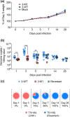
Analysis of MNV-3 VF1 virulence in vivo. C57BL/6 mice were orally gavaged with 1000 TCID50 of WT MNV-3 (3-WT) or MNV-3 M1 (3-M1). Mock inoculated mice were treated with DMEM. (a) Weights of mice are presented as relative to their day 0 weights (n=8). (b) vRNA extracted from faecal samples was quantified using qPCR. Points shaded blue represent VF1 expression (either from WT or revertant viruses). Red points represent absence of VF1 expression. Statistical analyses were calculated using two-way ANOVA with Bonferroni post-tests. (c) Percentages of mice that are shedding revertant viruses after acquiring the A5118G substitution. **P≤0.01; ***P≤0.001.
Fig. 5.
Fig. 5.
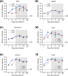
Replication kinetics in tissues. C57BL/6 mice were orally gavaged with 1000 TCID50 of WT MNV-3 (3-WT) or MNV-3 M1 (3-M1). Mock inoculated mice were treated with DMEM. vRNA was extracted from tissue samples, including (a) MLN, (b) spleen, (c) duodenum, (d) ileum, (e) caecum and (f) colon, and quantified using qPCR. Statistical analyses were carried out using an unpaired t-test. For WT MNV-3, n=20; for MNV-3 M1, n=20; and for mock, n=5. Shaded area shows the time points from which reversion was seen. *P≤0.05; **P≤0.01. LOD, limit of detection.
Fig. 6.
Fig. 6.
Role of MNV-3 VF1 in persistent infections. C57BL/6 mice were inoculated with WT MNV-3 (3-WT) or MNV-3 M3 (3-M3) at 1000 TCID50. Faecal samples were collected over a 3-month (84-day) period. (a) Infectious virus was isolated from faecal samples and titrated in RAW264.7 cells. Statistical analysis was carried out using two-way ANOVA with Bonferonni post-test (n=5). (b) Viral RNA from faecal samples was extracted and quantified with qPCR. Statistical analyses were carried out using a two-way ANOVA with Bonferroni post-test (n=5). *P≤0.05; **P≤0.01.

References

    1. Lopman BA, Steele D, Kirkwood CD, Parashar UD. The vast and varied global burden of norovirus: prospects for prevention and control. PLOS Med. 2016;13:e1001999. doi: 10.1371/journal.pmed.1001999. - DOI - PMC - PubMed
    1. Bányai K, Estes MK, Martella V, Parashar UD. Viral gastroenteritis. Lancet. 2018;392:175–186. doi: 10.1016/S0140-6736(18)31128-0. - DOI - PMC - PubMed
    1. Patel MM, Hall AJ, Vinjé J, Parashar UD. Noroviruses: A comprehensive review. J Clin Virol. 2009;44:1–8. doi: 10.1016/j.jcv.2008.10.009. - DOI - PubMed
    1. Ramani S, Kang G. Viruses causing childhood diarrhoea in the developing world. Curr Opin Infect Dis. 2009;22:477–482. doi: 10.1097/QCO.0b013e328330662f. - DOI - PubMed
    1. Karst SM, Wobus CE, Lay M, Davidson J, Virgin HW. STAT1-dependent innate immunity to a Norwalk-Like Virus. Science. 299:1575–1578. doi: 10.1126/science.1077905. - DOI - PubMed

Publication types

MeSH terms