. 2021 Sep 7;17(9):e1009285.
doi: 10.1371/journal.pcbi.1009285. eCollection 2021 Sep.

ASCENT (Automated Simulations to Characterize Electrical Nerve Thresholds): A pipeline for sample-specific computational modeling of electrical stimulation of peripheral nerves

Affiliations

ASCENT (Automated Simulations to Characterize Electrical Nerve Thresholds): A pipeline for sample-specific computational modeling of electrical stimulation of peripheral nerves

Eric D Musselman et al. PLoS Comput Biol. .

Abstract

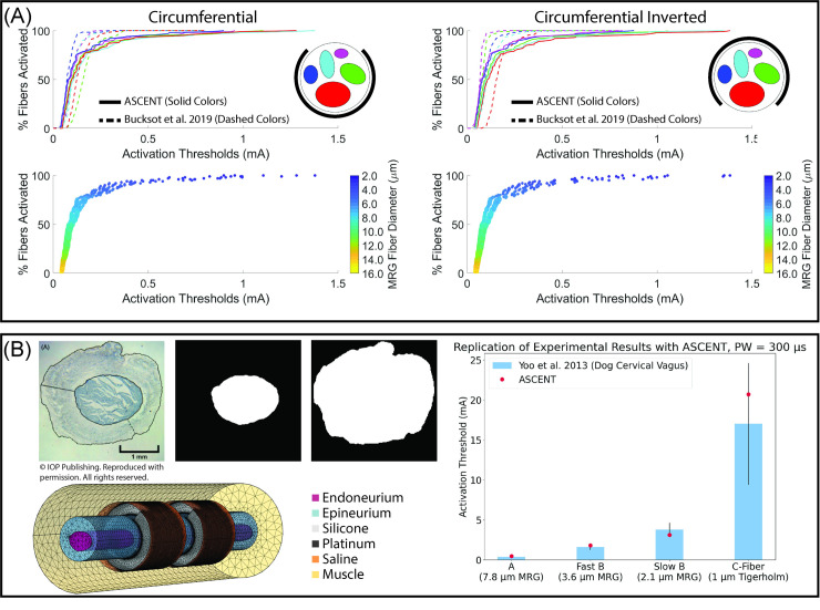
Electrical stimulation and block of peripheral nerves hold great promise for treatment of a range of disease and disorders, but promising results from preclinical studies often fail to translate to successful clinical therapies. Differences in neural anatomy across species require different electrodes and stimulation parameters to achieve equivalent nerve responses, and accounting for the consequences of these factors is difficult. We describe the implementation, validation, and application of a standardized, modular, and scalable computational modeling pipeline for biophysical simulations of electrical activation and block of nerve fibers within peripheral nerves. The ASCENT (Automated Simulations to Characterize Electrical Nerve Thresholds) pipeline provides a suite of built-in capabilities for user control over the entire workflow, including libraries for parts to assemble electrodes, electrical properties of biological materials, previously published fiber models, and common stimulation waveforms. We validated the accuracy of ASCENT calculations, verified usability in beta release, and provide several compelling examples of ASCENT-implemented models. ASCENT will enable the reproducibility of simulation data, and it will be used as a component of integrated simulations with other models (e.g., organ system models), to interpret experimental results, and to design experimental and clinical interventions for the advancement of peripheral nerve stimulation therapies.


Conflict of interest statement

The authors have declared that no competing interests exist.

Figures

Fig 1
Fig 1. Overview of the ASCENT pipeline for simulating the response of sample-specific nerves to electrical stimulation with custom cuff electrodes and stimulation waveforms.
FEM: finite element model.
Fig 2
Fig 2. Define sample-specific models with segmented nerve images.
We segmented the nerve boundary (n.tif), the perineurium (either “Combined” (c.tif), or both “Inners” (i.tif) and “Outers” (o.tif), or only “Inners”), and a horizontal “Scale Bar” (s.tif) of known length from a micrograph of a human cervical vagus nerve stained selectively for perineurium. In this example, two fascicles have multiple inner traces for their single outer trace (i.e., “peanut” fascicles), which requires their perineurium to be meshed; conversely, the other fascicles have a single inner trace for each outer trace, in which case the perineurium can be either meshed or modeled with a thin layer approximation. The nerve cross section is modeled in the (x,y,0) plane and extruded into the +z direction for the length of the nerve.
Fig 3
Fig 3. Parameterized preset cuff electrodes, modes to define fiber locations in nerve cross section, and modes to define conventional stimulation waveforms.
(A) Examples of “preset” cuffs commonly used in preclinical and clinical studies assembled using part primitives from S16 Text, which are provided in config/system/cuffs/. Custom cuff electrodes can be created from existing or new part primitives (S17 and S18 Text). (B) Types of FiberXYMode defining the (x, y)-coordinates of the fiber locations in the nerve cross section. The parameters needed to define the (x, y)-coordinates for each “FiberXYMode” are explained in S8 Text. (C) Parameterized definitions of conventional waveforms included in the ASCENT pipeline. Users can also define custom waveforms (S8 Text).
Fig 4
Fig 4. Modeling a multifascicular nerve with different cuff rotations to illustrate key ASCENT processes in context of configuration data hierarchy.
(A) Binary images of the fascicle boundaries (“inner” perineurium: i.tif) and nerve boundary (n.tif) to serve as inputs to the pipeline (left) and the resulting deformed traces for placement in a circular cuff electrode (right) following physics-based Python operations. The sample JSON configuration used is provided in examples/results/cuff_rotation/mock.json along with the binary images shown. (B) COMSOL FEM solved for the potentials generated by 1 mA delivered through the monopolar Enteromedics cuff. A contact impedance describes the perineurium ensheathing each fascicle (not shown). Given that it is a monopolar cuff, there is only one solution basis for a given cuff placement; a given Model will result in a basis solution for each contact, i.e., a basis *.mph file for each contact delivering 1 mA. (C) Inputs to NEURON simulations for activation and block: extracellular potentials (i.e., potentials/ sampled from bases/ corresponding to coordinates defined by fibersets/) and waveforms. The green arrow in the fibersets/ panel points to the (x,y,0)-location for the fiber potentials plotted along the length of the nerve in the potentials/ panel. (D) Heatmaps of activation and block thresholds for 10 μm diameter MRG fibers for four different cuff rotations on a multifascicular nerve. The black arc around the nerve represents the exposed contact length delivering current on the inside surface of the Enteromedics cuff. The heatmaps were generated using Query’s heatmaps() method.
Fig 5
Fig 5. Using ASCENT to replicate previously published computational models and to model an in vivo experiment.
(A) Recruitment curve for each fascicle in a model of the rabbit sciatic nerve for two rotations of a bipolar cuff electrode [11]. Fiber thresholds are in response to a 100 μs per phase biphasic rectangular pulse (i.e., BIPHASIC_PULSE_TRAIN). We simulated mammalian myelinated fibers at 0.5 μm diameter increments from 2 to 16 μm (i.e., MRG_INTERPOLATION) with 10 fibers of each diameter at consistent locations in each fascicle (i.e., UNIFORM_COUNT). We generated fiber recruitment curves by defining a population of fiber diameters from a normal distribution (8.85 ± 3.1 μm, 100 fibers per fascicle) and rounded each fiber diameter to the nearest 0.5 μm to draw from our modeled activation thresholds randomly. Configuration and input files used to replicate the results are provided in examples/results/bucksot_2019/. (B) Using ASCENT to model electrophysiology study of dog vagus stimulation [40]. We used Photoshop (Adobe Inc., San Jose, CA) to segment a micrograph from one of the animals in the original publication. We placed the nerve in a 2 mm diameter LivaNova helical cuff electrode delivering a 300 μs monophasic rectangular pulse (i.e., MONOPHASIC_PULSE_TRAIN), as used experimentally. We computed activation thresholds for myelinated (i.e., MRG_INTERPOLATION: 2.1, 3.6, and 7.8 μm diameter) and unmyelinated (i.e., TIGERHOLM: 1.0 μm diameter) fiber models, corresponding to the fiber types reported in the original study. We report the modeled threshold of a single fiber of each type at the centroid of the fascicle (i.e., CENTROID) (red dot) overlaid on the in vivo ENG thresholds (blue bars with black error bars: mean ± SD) of both myelinated (n = 5 dogs) and unmyelinated (n = 4 dogs) vagus nerve fibers. Our prior modeling studies show that fibers of a consistent diameter placed in the same fascicle have nearly identical thresholds [10]. Configuration and input files used to model the experiment are provided in examples/results/yoo_2013/.
Fig 6
Fig 6. Implementation of a human VNS model as studied in Arle et al. 2016 [39].
Top panel: segmented nerve geometry modeled in the original publication and image of FEM produced with ASCENT. As modeled in Arle et al. 2016 [39], the electrode lacks insulation. Bottom panel: heatmaps and recruitment curves for fiber activation thresholds across fiber diameters and pulse widths. We generated the heatmaps using Query’s heatmaps() method. Consistent with our prior modeling studies [10], the heatmaps show that fibers in the same fascicle with the same diameter have nearly identical thresholds. Configuration and input files used to replicate the results are provided in examples/results/arle_2016/.

Similar articles

Cited by

References

    1. Handforth A, DeGiorgio CM, Schachter SC, Uthman BM, Naritoku DK, Tecoma ES, et al.. Vagus nerve stimulation therapy for partial-onset seizures: a randomized active-control trial. Neurology. 1998Jul;51(1):48–55. Available from: doi: 10.1212/wnl.51.1.48 - DOI - PubMed
    1. Ben-Menachem E, Manon-Espaillat R, Ristanovic R, Wilder BJ, Stefan H, Mirza W, et al.. Vagus nerve stimulation for treatment of partial seizures: 1. A controlled study of effect on seizures. First International Vagus Nerve Stimulation Study Group. Epilepsia. 1994;35(3):616–26. Available from: doi: 10.1111/j.1528-1157.1994.tb02482.x - DOI - PubMed
    1. A randomized controlled trial of chronic vagus nerve stimulation for treatment of medically intractable seizures. The Vagus Nerve Stimulation Study Group. Neurology. 1995Feb;45(2):224–30. Available from: doi: 10.1212/wnl.45.2.224 - DOI - PubMed
    1. Marangell LB, Rush AJ, George MS, Sackeim HA, Johnson CR, Husain MM, et al.. Vagus nerve stimulation (VNS) for major depressive episodes: one year outcomes. Biol Psychiatry [Internet]. 2002. Feb 15;51(4):280–7. Available from: doi: 10.1016/s0006-3223(01)01343-9 - DOI - PubMed
    1. Nierenberg AA, Alpert JE, Gardner-Schuster EE, Seay S, Mischoulon D. Vagus Nerve Stimulation: 2-Year Outcomes for Bipolar Versus Unipolar Treatment-Resistant Depression. Biol Psychiatry [Internet]. 2008. Sep 15;64(6):455–60. Available from: doi: 10.1016/j.biopsych.2008.04.036 - DOI - PubMed

Publication types