

Megacystis in the first trimester of pregnancy: Prognostic factors and perinatal outcomes
- PMID: 34492029
- PMCID: PMC8423287
- DOI: 10.1371/journal.pone.0255890
Megacystis in the first trimester of pregnancy: Prognostic factors and perinatal outcomes
Abstract
Objective: To determine whether bladder size is associated with an unfavorable neonatal outcome, in the case of first-trimester megacystis.
Materials and methods: This was a retrospective observational study between 2009 and 2019 in two prenatal diagnosis centers. The inclusion criterion was an enlarged bladder (> 7 mm) diagnosed at the first ultrasound exam between 11 and 13+6 weeks of gestation. The main study endpoint was neonatal outcome based on bladder size. An adverse outcome was defined by the completion of a medical termination of pregnancy, the occurrence of in utero fetal death, or a neonatal death. Neonatal survival was considered as a favorable outcome and was defined by a live birth, with or without normal renal function, and with a normal karyotype.
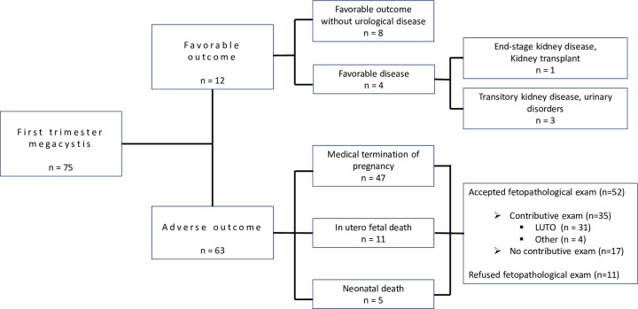
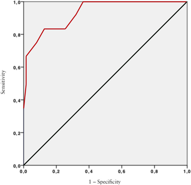
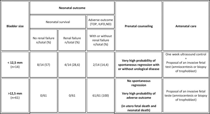
Results: Among 75 cases of first-trimester megacystis referred to prenatal diagnosis centers and included, there were 63 (84%) adverse outcomes and 12 (16%) live births. Fetuses with a bladder diameter of less than 12.5 mm may have a favorable outcome, with or without urological problems, with a high sensitivity (83.3%) and specificity (87.3%), area under the ROC curve = 0.93, 95% CI (0.86-0.99), p< 0.001. Fetal autopsy was performed in 52 (82.5%) cases of adverse outcome. In the 12 cases of favorable outcome, pediatric follow-up was normal and non-pathological in 8 (66.7%).
Conclusion: Bladder diameter appears to be a predictive marker for neonatal outcome. Fetuses with smaller megacystis (7-10 mm) have a significantly higher chance of progressing to a favorable outcome. Urethral stenosis and atresia are the main diagnoses made when first-trimester megacystis is observed. Karyotyping is important regardless of bladder diameter.
Conflict of interest statement
The authors have declared that no competing interests exist.
Figures
Similar articles
-
Fetal megacystis: prediction of spontaneous resolution and outcome.Ultrasound Obstet Gynecol. 2017 Oct;50(4):458-463. doi: 10.1002/uog.17422. Epub 2017 Sep 5. Ultrasound Obstet Gynecol. 2017. PMID: 28133847
-
Prenatal detection of megacystis: not always an adverse prognostic factor. Experience in 25 consecutive cases in a tertiary referral center, with complete neonatal outcome and follow-up.J Pediatr Urol. 2017 Oct;13(5):486.e1-486.e10. doi: 10.1016/j.jpurol.2017.04.001. Epub 2017 Apr 14. J Pediatr Urol. 2017. PMID: 28495235
-
Early fetal megacystis: Is it possible to predict the prognosis in the first trimester?J Perinat Med. 2018 Nov 27;46(9):1035-1039. doi: 10.1515/jpm-2017-0351. J Perinat Med. 2018. PMID: 29369818
-
Fetal megacystis: A systematic review.J Pediatr Urol. 2017 Feb;13(1):7-15. doi: 10.1016/j.jpurol.2016.09.003. Epub 2016 Oct 8. J Pediatr Urol. 2017. PMID: 27889224
-
Outcomes in fetuses diagnosed with megacystis: Systematic review and meta-analysis.Eur J Obstet Gynecol Reprod Biol. 2019 Feb;233:120-126. doi: 10.1016/j.ejogrb.2018.12.007. Epub 2018 Dec 13. Eur J Obstet Gynecol Reprod Biol. 2019. PMID: 30594021
Cited by
-
Single-center outcome analysis of 46 fetuses with megacystis after intrauterine vesico-amniotic shunting with the Somatex®intrauterine shunt.Arch Gynecol Obstet. 2024 Jan;309(1):145-158. doi: 10.1007/s00404-022-06905-6. Epub 2023 Jan 5. Arch Gynecol Obstet. 2024. PMID: 36604332 Free PMC article.
-
Obstructive or non-obstructive megacystis: a prenatal dilemma.Front Pediatr. 2024 Jul 2;12:1379267. doi: 10.3389/fped.2024.1379267. eCollection 2024. Front Pediatr. 2024. PMID: 39015208 Free PMC article.
References
-
- Karim JN, Roberts NW, Salomon LJ, Papageorghiou AT. Systematic review of first-trimester ultrasound screening for detection of fetal structural anomalies and factors that affect screening performance. Ultrasound Obstet Gynecol Off J Int Soc Ultrasound Obstet Gynecol. 2017Oct;50(4):429–41. - PubMed
Publication types
MeSH terms
Supplementary concepts
LinkOut - more resources
Full Text Sources
Medical
