

Migrasome biogenesis and functions
- PMID: 34492154
- PMCID: PMC9786993
- DOI: 10.1111/febs.16183
Migrasome biogenesis and functions
Abstract
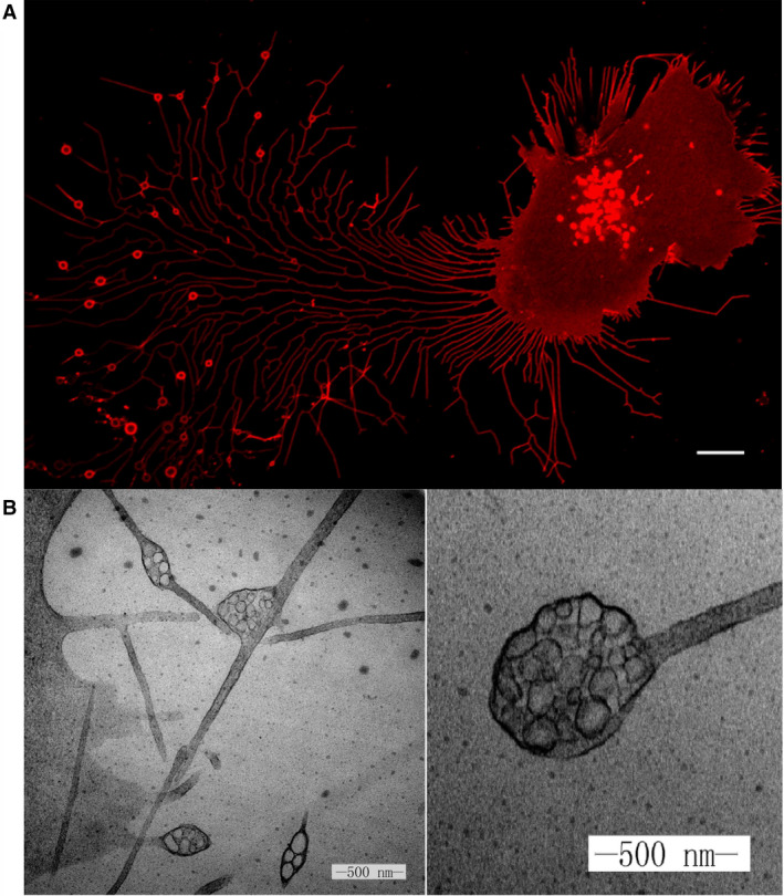
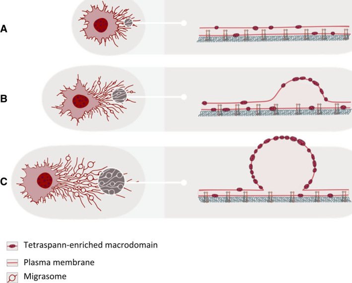
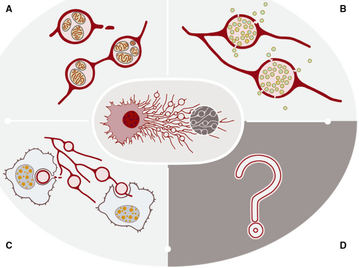
The migrasome is a newly discovered organelle produced by migrating cells. As cells migrate, long and thin retraction fibers are left in their wake. On these fibers, we discovered the production of a pomegranate-like structure, which we named migrasomes. The production of migrasomes is highly correlated with the migration of cells. Currently, it has been demonstrated the migrasomes exhibit three modes of action: release of signaling molecules through rupturing or leaking, carriers of damaged mitochondria, and lateral transfer of mRNA or proteins. In this review, we would like to discuss, in detail, the functions, mechanisms, and potential applications of this newly discovered cell organelle.
Keywords: cell migration; confocal microscopy; migrasomes; tetraspanin; transmission electron microscopy.
© 2021 The Authors. The FEBS Journal published by John Wiley & Sons Ltd on behalf of Federation of European Biochemical Societies.
Conflict of interest statement
The authors declare no conflict of interest and agree on the submission an publication of this manuscript.
Figures
Similar articles
-
Detection, Purification, Characterization, and Manipulation of Migrasomes.Curr Protoc. 2023 Aug;3(8):e856. doi: 10.1002/cpz1.856. Curr Protoc. 2023. PMID: 37540780
-
Differences between migrasome, a 'new organelle', and exosome.J Cell Mol Med. 2023 Dec;27(23):3672-3680. doi: 10.1111/jcmm.17942. Epub 2023 Sep 4. J Cell Mol Med. 2023. PMID: 37665060 Free PMC article. Review.
-
Tetraspanin 4 stabilizes membrane swellings and facilitates their maturation into migrasomes.Nat Commun. 2023 Feb 23;14(1):1037. doi: 10.1038/s41467-023-36596-9. Nat Commun. 2023. PMID: 36823145 Free PMC article.
-
Migrasome: a novel insight into unraveling physiological and pathological function.Mol Biol Rep. 2025 May 28;52(1):509. doi: 10.1007/s11033-025-10615-y. Mol Biol Rep. 2025. PMID: 40434508 Review.
-
Biophysical aspects of migrasome organelle formation and their diverse cellular functions.Bioessays. 2024 Aug;46(8):e2400051. doi: 10.1002/bies.202400051. Epub 2024 Jun 23. Bioessays. 2024. PMID: 38922978 Free PMC article. Review.
Cited by
-
Extracellular Vesicles and Membrane Protrusions in Developmental Signaling.J Dev Biol. 2022 Sep 21;10(4):39. doi: 10.3390/jdb10040039. J Dev Biol. 2022. PMID: 36278544 Free PMC article. Review.
-
Heterogeneity of Extracellular Vesicles and Particles: Molecular Voxels in the Blood Borne "Hologram" of Organ Function, Disfunction and Cancer.Arch Immunol Ther Exp (Warsz). 2023 Feb 2;71(1):5. doi: 10.1007/s00005-023-00671-2. Arch Immunol Ther Exp (Warsz). 2023. PMID: 36729313 Review.
-
Novel insights into the roles of migrasome in cancer.Discov Oncol. 2024 May 15;15(1):166. doi: 10.1007/s12672-024-00942-0. Discov Oncol. 2024. PMID: 38748047 Free PMC article. Review.
-
Contribution of extracellular vesicles for the pathogenesis of retinal diseases: shedding light on blood-retinal barrier dysfunction.J Biomed Sci. 2024 May 10;31(1):48. doi: 10.1186/s12929-024-01036-3. J Biomed Sci. 2024. PMID: 38730462 Free PMC article. Review.
-
Exomeres and supermeres: Current advances and perspectives.Bioact Mater. 2025 Apr 16;50:322-343. doi: 10.1016/j.bioactmat.2025.04.012. eCollection 2025 Aug. Bioact Mater. 2025. PMID: 40276541 Free PMC article. Review.
References
-
- Yu L (2021) Migrasomes: the knowns, the known unknowns and the unknown unknowns: a personal perspective. Sci China Life Sci 64, 162–166. - PubMed
Publication types
MeSH terms
LinkOut - more resources
Full Text Sources
Other Literature Sources
Research Materials
