

Shigella ubiquitin ligase IpaH7.8 targets gasdermin D for degradation to prevent pyroptosis and enable infection
- PMID: 34492225
- PMCID: PMC9122893
- DOI: 10.1016/j.chom.2021.08.010
Shigella ubiquitin ligase IpaH7.8 targets gasdermin D for degradation to prevent pyroptosis and enable infection
Abstract
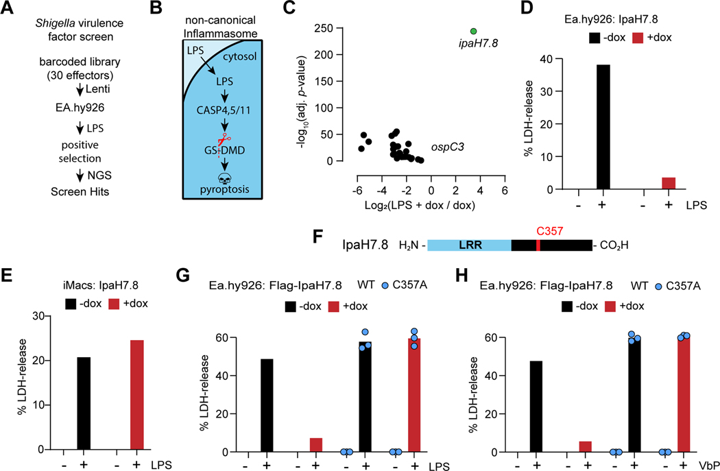
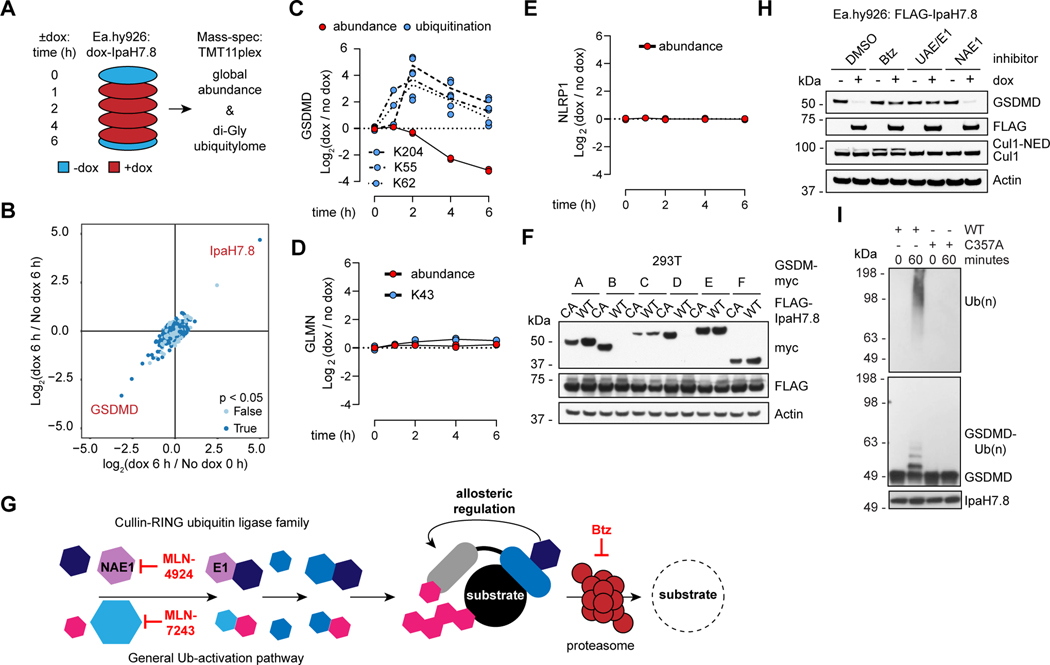
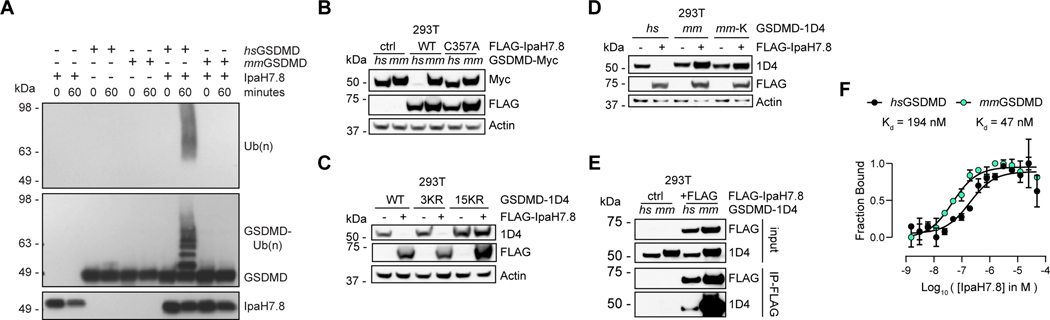
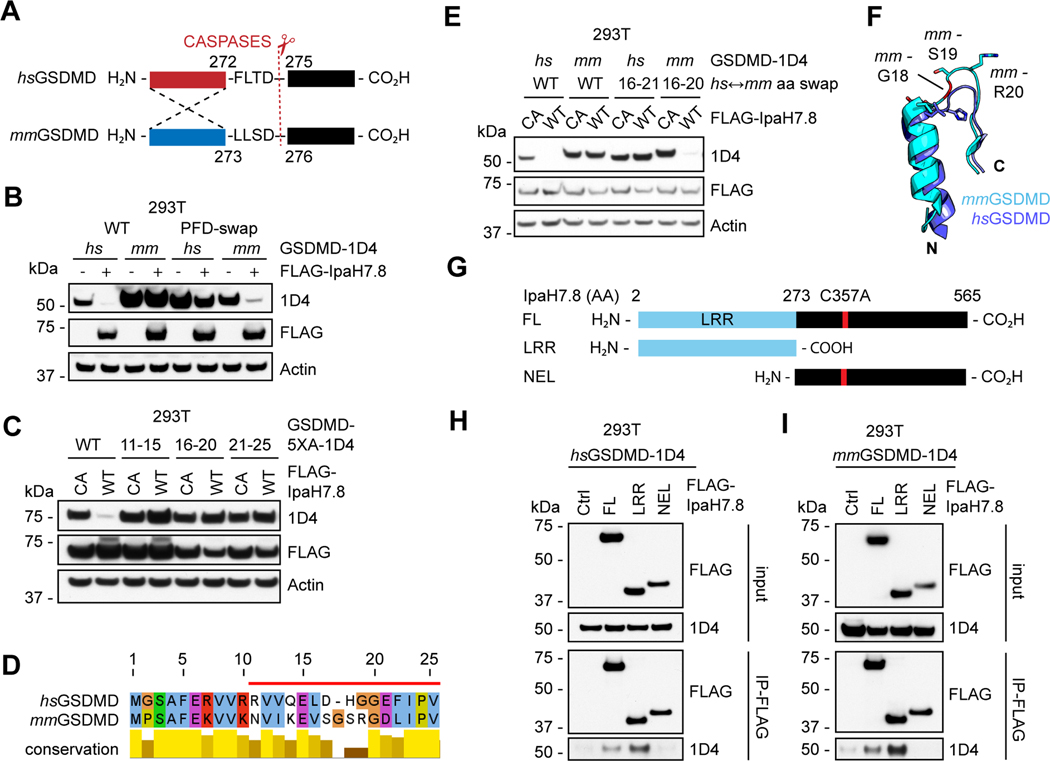
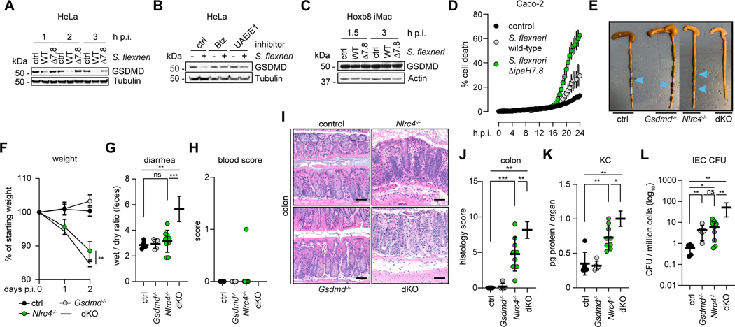
The pore-forming protein gasdermin D (GSDMD) executes lytic cell death called pyroptosis to eliminate the replicative niche of intracellular pathogens. Evolution favors pathogens that circumvent this host defense mechanism. Here, we show that the Shigella ubiquitin ligase IpaH7.8 functions as an inhibitor of GSDMD. Shigella is an enteroinvasive bacterium that causes hemorrhagic gastroenteritis in primates, but not rodents. IpaH7.8 contributes to species specificity by ubiquitinating human, but not mouse, GSDMD and targeting it for proteasomal degradation. Accordingly, infection of human epithelial cells with IpaH7.8-deficient Shigella flexneri results in increased GSDMD-dependent cell death compared with wild type. Consistent with pyroptosis contributing to murine disease resistance, eliminating GSDMD from NLRC4-deficient mice, which are already sensitized to oral infection with Shigella flexneri, leads to further enhanced bacterial replication and increased disease severity. This work highlights a species-specific pathogen arms race focused on maintenance of host cell viability.
Keywords: GSDMD; Shigella; inflammasome; proteasome; pyroptosis; ubiquitin ligase; virulence.
Copyright © 2021 Genentech. Published by Elsevier Inc. All rights reserved.
Conflict of interest statement
Declaration of interests All authors except J.R., R.A.C., and R.E.V. are employees of Genentech. R.E.V. is an investigator of the Howard Hughes Medical Institute and a consultant for Ventus Therapeutics and Tempest Therapeutics.
Figures
Comment in
-
Shigella shuts down the pyrop-technic show.Cell Host Microbe. 2021 Oct 13;29(10):1473-1476. doi: 10.1016/j.chom.2021.09.012. Cell Host Microbe. 2021. PMID: 34648738
Similar articles
-
Shigella IpaH7.8 E3 ubiquitin ligase targets glomulin and activates inflammasomes to demolish macrophages.Proc Natl Acad Sci U S A. 2014 Oct 7;111(40):E4254-63. doi: 10.1073/pnas.1324021111. Epub 2014 Sep 22. Proc Natl Acad Sci U S A. 2014. PMID: 25246571 Free PMC article.
-
Pathogenic ubiquitination of GSDMB inhibits NK cell bactericidal functions.Cell. 2021 Jun 10;184(12):3178-3191.e18. doi: 10.1016/j.cell.2021.04.036. Epub 2021 May 21. Cell. 2021. PMID: 34022140 Free PMC article.
-
Insights into the GSDMB-mediated cellular lysis and its targeting by IpaH7.8.Nat Commun. 2023 Jan 4;14(1):61. doi: 10.1038/s41467-022-35725-0. Nat Commun. 2023. PMID: 36599845 Free PMC article.
-
Regulation of Lytic and Non-Lytic Functions of Gasdermin Pores.J Mol Biol. 2022 Feb 28;434(4):167246. doi: 10.1016/j.jmb.2021.167246. Epub 2021 Sep 17. J Mol Biol. 2022. PMID: 34537232 Review.
-
Chemical Modulation of Gasdermin-Mediated Pyroptosis and Therapeutic Potential.J Mol Biol. 2022 Feb 28;434(4):167183. doi: 10.1016/j.jmb.2021.167183. Epub 2021 Aug 3. J Mol Biol. 2022. PMID: 34358546 Free PMC article. Review.
Cited by
-
No longer married to inflammasome signaling: the diverse interacting pathways leading to pyroptotic cell death.Biochem J. 2022 May 27;479(10):1083-1102. doi: 10.1042/BCJ20210711. Biochem J. 2022. PMID: 35608339 Free PMC article. Review.
-
Palmitoylation at a conserved cysteine residue facilitates gasdermin D-mediated pyroptosis and cytokine release.Proc Natl Acad Sci U S A. 2024 Jul 16;121(29):e2400883121. doi: 10.1073/pnas.2400883121. Epub 2024 Jul 9. Proc Natl Acad Sci U S A. 2024. PMID: 38980908 Free PMC article.
-
From gum inflammation to oral cancers: pyroptosis as the molecular torchbearer in periodontitis-driven carcinogenesis.Discov Oncol. 2025 Sep 1;16(1):1663. doi: 10.1007/s12672-025-03508-w. Discov Oncol. 2025. PMID: 40888849 Free PMC article. Review.
-
Gasdermin D-Mediated Pyroptosis Exerts Two Opposite Effects of Resisting Enzymatic Digestion and Expanding Inflammatory Response in Acute Pancreatitis.Adv Sci (Weinh). 2025 Aug;12(31):e02412. doi: 10.1002/advs.202502412. Epub 2025 May 29. Adv Sci (Weinh). 2025. PMID: 40439590 Free PMC article.
-
Pyroptosis in periodontitis: From the intricate interaction with apoptosis, NETosis, and necroptosis to the therapeutic prospects.Front Cell Infect Microbiol. 2022 Aug 16;12:953277. doi: 10.3389/fcimb.2022.953277. eCollection 2022. Front Cell Infect Microbiol. 2022. PMID: 36093182 Free PMC article. Review.
References
-
- Brownell JE, Sintchak MD, Gavin JM, Liao H, Bruzzese FJ, Bump NJ, Soucy TA, Milhollen MA, Yang X, Burkhardt AL, Ma J, Loke HK., Lingaraj T, Wu D, Hamman KB, Spelman JL, Cullis CA, Langston SP, Vyskocil S, Sells TB, Mallender WD, Visiers I, Li P, Claiborne CF, Rolfe M, Bolen JB, Rick LR (2010) Substrate-assisted inhibition of ubiquitin-like protein-activating enzymes: the nedd8 E1 inhibitor MLN4924 forms a NEDD8-AMP mimetic in situ. Mol. Cell 37, 102–111. - PubMed
MeSH terms
Substances
Grants and funding
LinkOut - more resources
Full Text Sources
Other Literature Sources
Molecular Biology Databases
