. 2021 Dec 15:194:113565.
doi: 10.1016/j.bios.2021.113565. Epub 2021 Aug 13.

Flow radiocytometry using droplet optofluidics

Affiliations

Flow radiocytometry using droplet optofluidics

Byunghang Ha et al. Biosens Bioelectron. .

Abstract

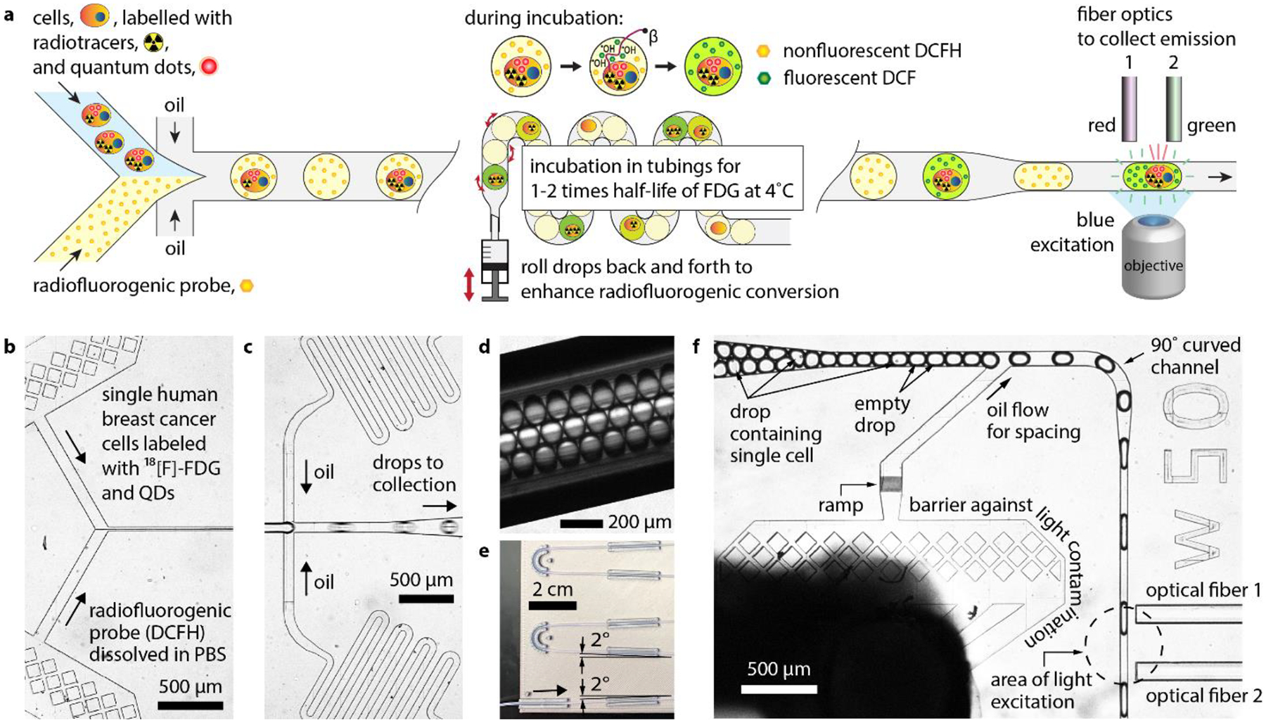
Flow-based cytometry methods are widely used to analyze heterogeneous cell populations. However, their use for small molecule studies remains limited due to bulky fluorescent labels that often interfere with biochemical activity in cells. In contrast, radiotracers require minimal modification of their target molecules and can track biochemical processes with negligible interference and high specificity. Here, we introduce flow radiocytometry (FRCM) that broadens the scope of current cytometry methods to include beta-emitting radiotracers as probes for single cell studies. FRCM uses droplet microfluidics and radiofluorogenesis to translate the radioactivity of single cells into a fluorescent signal that is then read out using a high-throughput optofluidic device. As a proof of concept, we quantitated [18F]fluorodeoxyglucose radiotracer uptake in single human breast cancer cells and successfully assessed the metabolic flux of glucose and its heterogeneity at the cellular level. We believe FRCM has potential applications ranging from analytical assays for cancer and other diseases to development of small-molecule drugs.

Keywords: Droplet microfluidics; Fluorodeoxyglucose; Optofluidics; Radiochemistry; Radiofluorogenesis; Single-cell analysis.


Conflict of interest statement

COMPETING INTERESTS

G.P. is listed as inventor on a patent (US 20160025701 A1) related to this work. Other authors declare no conflict of interest.

Declaration of interests

The authors declare the following financial interests/personal relationships which may be considered as potential competing interests:

G.P. is listed as inventor on a patent (US 20160025701 A1) related to this work. Other authors declare no conflict of interest.

Figures

Figure 1.
Figure 1.
(a) Molecular structures of D-glucose, fluorine-18-radiolabeled glucose analog (2-deoxy-2-[18F]-fluoro-D-glucose or [18F]FDG), and fluorescent glucose analog (2-NBDG). [14C]glucose is made by replacing one of the carbon atom of a D-glucose molecule with its radioisotope 14C. (b) Illustration showing production of ROS from radioactive beta decay. Proton (P) decays into neutron (N), emitting positron (e+) and neutrino (v). The emitted positron travels a finite distance, interacting with water molecules along its path and creating ROS, until it annihilates with an electron (e−), resulting in two antiparallel 511 keV photons. (c) Illustration showing principle of radiofluorogenic conversion. A single radioactive cell is encapsulated into a water-in-oil drop. As the radiotracer decays, the emitted beta radiation creates ROS such as hydroxyl radicals, which in turn mediate the radiofluorogenic conversion. (d) Nonfluorescent dichlorodihydrofluorescein (DCFH) is converted into fluorescent dichlorofluorescein (DCF) after losing two protons from interacting with radiation-induced ROS.
Figure 2.
Figure 2.
Device overview and design. (a) Schematics showing drop generation, incubation, and read. (b) Optical micrograph (top view) showing cell samples and radiofluorogenic probe solution being introduced and mixed in DropGen microchip (c) Optical micrograph showing the cross-junction microfluidic channels where the mixture (i.e., dispersed phase) including the cells was encapsulated in water-in-oil drops. (d) Optical micrograph of polytetrafluoroethylene tubing where drops were incubated. (e) Picture of the tubing and holder. (f) Optical micrograph of DropRead microchip in which drops were introduced to have their fluorescence measured after incubation.
Figure 3.
Figure 3.
Optofluidic configuration and measurement. (a) Schematic showing the optofluidic configuration. (b) The two PMT signals as a function of elapsed time.
Figure 4.
Figure 4.
Kinetic fluorescence spectroscopy measurements of (a) DCFH solutions and (b) mixture of DCFH and FDG. (c) Signal to background ratio (SBR) as a function of elapsed time. (d) SBR measured at increasing elapsed times as a function of the ratio of radioactivity to DCFH concentration.
Figure 5.
Figure 5.
Fluorescence measurements of drops containing non-radiolabeled cells (control) vs. radiolabeled cells (sample). (a,b) Micrographs of drops at the inlet of DropRead microchips: (a) control and (b) sample; bright field image on the left and FITC fluorescence image on the right. (c,d) PMT measurements as a function of time for (c) control and (d) sample assays.
Figure 6.
Figure 6.
(a) Western blot comparing SLC2A1 knockdown (shGLUT1) to scrambled shRNA control (shSCR) in MDA-MB-231 cells. (b) Averaged radioactivity of shSCR and shGLUT1 samples measured using bulk dose calibrator and hemocytometry. The error bars show standard deviation and associated with cell counting. (c) Half-violin plot summarizing the drop read results. Each dot in the scatter plots on the left-hand side represents a single drop measured by FRCM. The frequency distribution corresponding to the scatter plot is shown on the right-hand side. Grey: non-radioactive cell sample (i.e., control), red: FDG-labeled control shSCR cell sample, and blue: FDG-labeled shGLUT1 cell sample.

References

    1. Cai L, Friedman N, Xie XS, 2006. Nature 440, 358–362. - PubMed
    1. Cantelmo AR, Conradi L-C, Brajic A, Goveia J, Kalucka J, Pircher A, Chaturvedi P, Hol J, Thienpont B, Teuwen L-A, 2016. Cancer cell 30, 968–985. - PMC - PubMed
    1. Chattopadhyay A, Shrivastava S, Chaudhuri A, 2016., John Wiley & Sons, Inc
    1. Chen X, Zhong Z, Xu Z, Chen L, Wang Y, 2010. Free radical research 44, 587–604. - PubMed
    1. Conti M, Eriksson L, 2016. EJNMMI physics 3, 8. - PMC - PubMed