. 2021 Sep 21;10(18):e017524.
doi: 10.1161/JAHA.120.017524. Epub 2021 Sep 8.

Cysteamine Decreases Low-Density Lipoprotein Oxidation, Causes Regression of Atherosclerosis, and Improves Liver and Muscle Function in Low-Density Lipoprotein Receptor-Deficient Mice

Affiliations

Cysteamine Decreases Low-Density Lipoprotein Oxidation, Causes Regression of Atherosclerosis, and Improves Liver and Muscle Function in Low-Density Lipoprotein Receptor-Deficient Mice

Feroz Ahmad et al. J Am Heart Assoc. .

Abstract

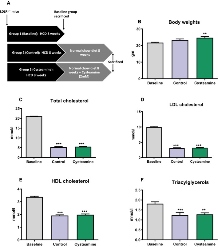
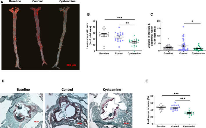
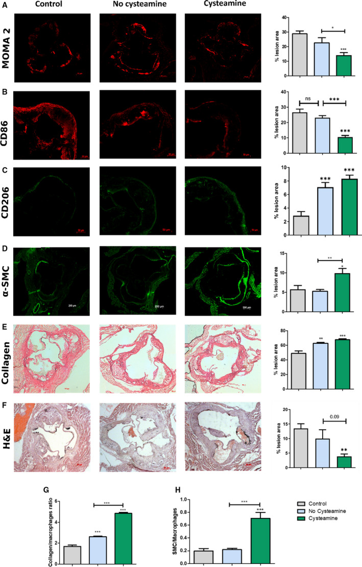
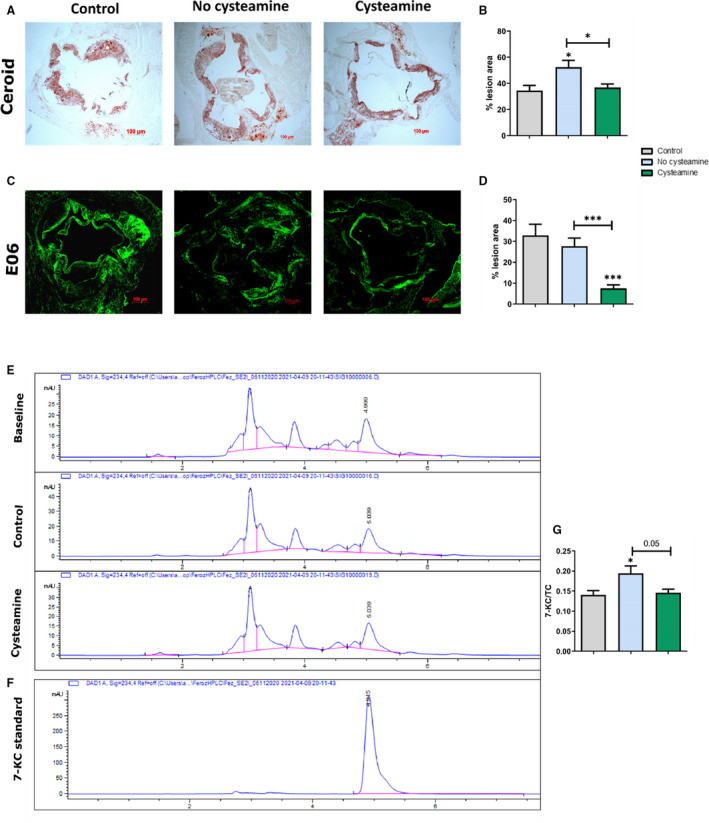
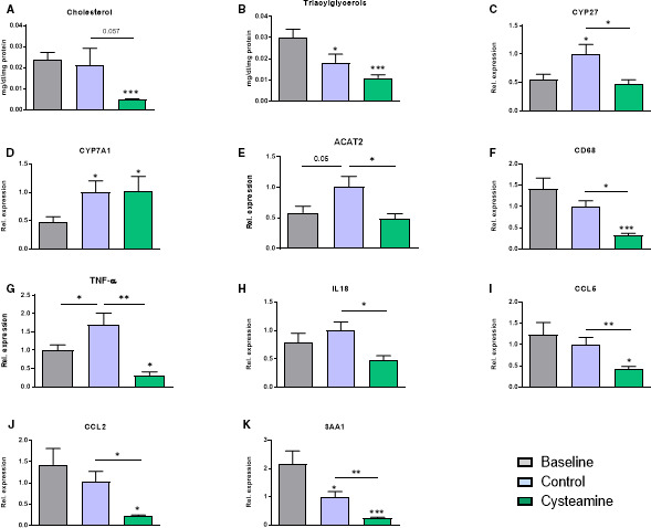
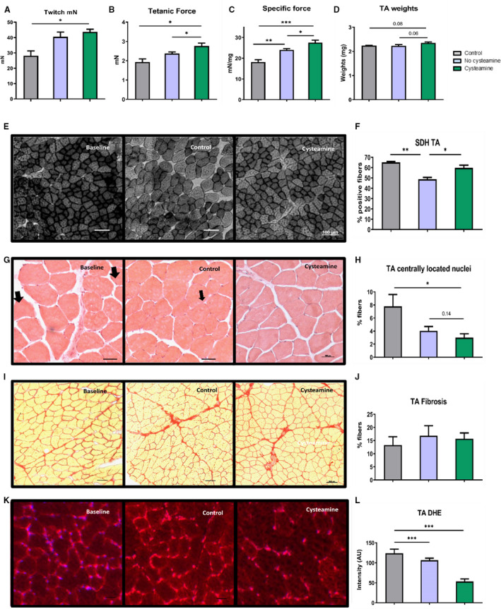
Background We have shown previously that low-density lipoprotein (LDL) can be oxidized in the lysosomes of macrophages, that this oxidation can be inhibited by cysteamine, an antioxidant that accumulates in lysosomes, and that this drug decreases atherosclerosis in LDL receptor-deficient mice fed a high-fat diet. We have now performed a regression study with cysteamine, which is of more relevance to the treatment of human disease. Methods and Results LDL receptor-deficient mice were fed a high-fat diet to induce atherosclerotic lesions. They were then reared on chow diet and drinking water containing cysteamine or plain drinking water. Aortic atherosclerosis was assessed, and samples of liver and skeletal muscle were analyzed. There was no regression of atherosclerosis in the control mice, but cysteamine caused regression of between 32% and 56% compared with the control group, depending on the site of the lesions. Cysteamine substantially increased markers of lesion stability, decreased ceroid, and greatly decreased oxidized phospholipids in the lesions. The liver lipid levels and expression of cluster of differentiation 68, acetyl-coenzyme A acetyltransferase 2, cytochromes P450 (CYP)27, and proinflammatory cytokines and chemokines were decreased by cysteamine. Skeletal muscle function and oxidative fibers were increased by cysteamine. There were no changes in the plasma total cholesterol, LDL cholesterol, high-density lipoprotein cholesterol, or triacylglycerol concentrations attributable to cysteamine. Conclusions Inhibiting the lysosomal oxidation of LDL in atherosclerotic lesions by antioxidants targeted at lysosomes causes the regression of atherosclerosis and improves liver and muscle characteristics in mice and might be a promising novel therapy for atherosclerosis in patients.

Keywords: antioxidant; atherosclerosis; lipoprotein; low‐density lipoprotein.


Conflict of interest statement

None.

Figures

Figure 1
Figure 1. Effect of cysteamine on body weights and plasma lipids in low‐density lipoprotein (LDL) receptor–knockout mice.
A, Plan of the study. Sixty female LDL receptor–deficient (LDLr−/−) mice were divided into 3 groups and fed a high‐cholesterol diet (HCD) for 8 weeks to induce atherosclerosis. One group was euthanized for baseline measurements. The remaining 40 mice were switched to a normal chow diet and divided into 2 groups to receive no cysteamine (control) or cysteamine at 2.2 mmol/L in drinking water for 8 weeks. Body weight of the mice and plasma lipids were measured at the end of the study. Cysteamine has no effect on the body weights (B); the levels of plasma lipids (CF) were reduced after switching the mice to a normal chow diet, and cysteamine had no effect on them. There were 17 to 20 mice in each group, and the horizontal line shows the group mean±SEM. Data were analyzed by ANOVA, followed by the Tukey post hoc test. HDL indicates high‐density lipoprotein. **P<0.001 vs the baseline group, ***P<0.0001 vs baseline group.
Figure 2
Figure 2. Cysteamine reduced existing atherosclerosis in low‐density lipoprotein receptor–deficient mice.
A, Representative images to show atherosclerotic lesions in the aorta of baseline, control, and mice treated with cysteamine (2.2 mmol/L in drinking water) stained with Oil Red O. Bar=500 μm. Data points show lesion areas in individual mice in each group in the aortic arch (B) and the rest of the thoracic plus abdominal aorta (C) of baseline, control, and cysteamine‐treated mice. Representative images to show atherosclerotic lesions in the aortic root (D) and lesion areas in individual mice (E). There were 17 to 20 mice in each group, and the horizontal line shows the group mean±SEM. Data were analyzed by ANOVA, followed by the Tukey post hoc test. *P<0.05, **P<0.01, ***P<0.001.
Figure 3
Figure 3. Effect of cysteamine on atherosclerotic plaque composition.
Sections from the aortic root were stained for monocytes/macrophages, cluster of differentiation (CD) 86, CD206, smooth muscle cells (SMCs), collagen, and hematoxylin and eosin (H&E), and the staining was quantified. Quantitation and representative photomicrographs of anti‐monocyte+macrophage antibody (MOMA‐2) (monocytes/macrophages) (A), CD86 (B), CD206 (C), actin (SMCs) (D), Picrosirius Red (collagen) staining (E) and H&E (F) in transverse sections from the aortic root of baseline, control, and cysteamine‐treated mice and percentage of acellular area. G and H, The ratio of collagen/macrophage areas and ratio of SMCs/macrophage areas were calculated as markers of lesion stability. Horizontal bars represent the group mean±SEM for 10 to 12 mice in each group. Data were analyzed by ANOVA, followed by the Tukey post hoc test. α‐SMC indicates α‐smooth muscle actin. *P<0.05, **P<0.01, ***P<0.001.
Figure 4
Figure 4. Effect of cysteamine on lipoprotein oxidation in aortic lesions.
A and B, Quantitation and representative images to show ceroid levels in the atherosclerotic lesions. C and D, Quantitation and representative images to show immunofluorescence staining with the E06 monoclonal antibody against oxidized phospholipids. E, Representative high‐performance liquid chromatography chromatograms of aortic root lesions of baseline, control, and cysteamine‐treated mice. F, Chromatogram of 10 µmol/L 7‐ketocholesterol (7‐KC) standard at 234 nm. G, Ratio of 7‐KC/total cholesterol (TC) in the aortic lesions. There were 17 to 20 mice in each group, and the horizontal line shows the group mean±SEM. Data were analyzed by ANOVA, followed by the Tukey post hoc test. *P<0.05, ***P<0.001.
Figure 5
Figure 5. Effect of cysteamine on hepatic cholesterol levels and inflammation.
The mean total hepatic cholesterol (A) and triacylglycerol (B) levels were reduced by cysteamine compared with the baseline mice. Total RNA was isolated from mouse liver, and gene expression of various cholesterol‐metabolizing enzymes and proinflammatory proteins was measured. The increased expression of CYP 27 and ACAT2 (acetyl–coenzyme A acetyltransferase) in the control mice was reduced by cysteamine (C and E). CYP7A1 expression was elevated during the regression phase but was not affected by cysteamine (D). Cysteamine treatment reduced the expression of hepatic proinflammatory markers: cluster of differentiation (CD) 68, tumor necrosis factor (TNF)‐α, interleukin (IL)‐18, CCL5, CCL2, and serum amyloid A1 (SAA1) (FK). Horizontal bars represent group the mean±SEM for 17 to 20 mice in each group. Data were analyzed by ANOVA, followed by the Tukey post hoc test. Rel. indicates relative. *P<0.05, **P<0.01, ***P<0.001.
Figure 6
Figure 6. Effect of cysteamine on skeletal muscles.
Muscle contraction was measured through ex vivo assessment of tibialis anterior (TA) twitch (A), tetanic force (B), and specific force (C). D, TA weights normalized to body weights. E, Qualitative changes in muscle function were evaluated by staining for succinate dehydrogenase (SDH). F, Quantification of stained SDH fibers. Hematoxylin and eosin–stained sections (G) were used to see muscle regeneration by calculating the percentage of fibers with centrally located nuclei (arrows) (H). I and J, Muscle fibrosis was calculated by staining for collagen deposition (Picrosirius Red) in TA. DHE‐stained sections (K) and quantification of DHE (L) are shown. Horizontal bars represent group mean±SEM for 17 to 20 mice in each group. Data were analyzed by ANOVA, followed by the Tukey post hoc test. *P<0.05, **P<0.01, ***P<0.001.

References

    1. Libby P, Hansson GK. From focal lipid storage to systemic inflammation: JACC review topic of the week. J Am Coll Cardiol. 2019;74:1594–1607. DOI: 10.1016/j.jacc.2019.07.061. - DOI - PMC - PubMed
    1. Remmerie A, Scott CL. Macrophages and lipid metabolism. Cell Immunol. 2018;330:27–42. DOI: 10.1016/j.cellimm.2018.01.020. - DOI - PMC - PubMed
    1. Fatkhullina AR, Peshkova IO, Koltsova EK. The role of cytokines in the development of atherosclerosis. Biochem (Mosc). 2016;81:1358–1370. DOI: 10.1134/S0006297916110134. - DOI - PMC - PubMed
    1. Ridker PM, Everett BM, Thuren T, MacFadyen JG, Chang WH, Ballantyne C, Fonseca F, Nicolau J, Koenig W, Anker SD, et al. Antiinflammatory therapy with canakinumab for atherosclerotic disease. N Engl J Med. 2017;377:1119–1131. DOI: 10.1056/NEJMoa1707914. - DOI - PubMed
    1. Kleemann R, Verschuren L, van Erk MJ, Nikolsky Y, Cnubben NHP, Verheij ER, Smilde AK, Hendriks HFJ, Zadelaar S, Smith GJ, et al. Atherosclerosis and liver inflammation induced by increased dietary cholesterol intake: a combined transcriptomics and metabolomics analysis. Genome Biol. 2007;8:R200. DOI: 10.1186/gb-2007-8-9-r200. - DOI - PMC - PubMed

Publication types

LinkOut - more resources