Review
. 2021 Sep;11(9):210189.
doi: 10.1098/rsob.210189. Epub 2021 Sep 8.

Recent insights into mechanisms preventing ectopic centromere formation

Affiliations
Review

Recent insights into mechanisms preventing ectopic centromere formation

Qianhua Dong et al. Open Biol. 2021 Sep.

Abstract

The centromere is a specialized chromosomal structure essential for chromosome segregation. Centromere dysfunction leads to chromosome segregation errors and genome instability. In most eukaryotes, centromere identity is specified epigenetically by CENP-A, a centromere-specific histone H3 variant. CENP-A replaces histone H3 in centromeres, and nucleates the assembly of the kinetochore complex. Mislocalization of CENP-A to non-centromeric regions causes ectopic assembly of CENP-A chromatin, which has a devastating impact on chromosome segregation and has been linked to a variety of human cancers. How non-centromeric regions are protected from CENP-A misincorporation in normal cells is largely unexplored. Here, we review the most recent advances on the mechanisms underlying the prevention of ectopic centromere formation, and discuss the implications in human disease.

Keywords: CENP-A; chromosomes segregation; ectopic centromeres; epigenetics; kinetochore; neocentromere.


Conflict of interest statement

We declare we have no interests to declare.

Figures

Figure 1.
Figure 1.
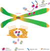
Formation of ectopic centromeres can lead to chromosome segregation errors. Normal centromeres ensure equal segregation of chromosomes into daughter nuclei. Unequal chromosome segregation, chromosome breakage and lagging chromosomes can be observed when cells fail to prevent the formation of ectopic centromeres.
Figure 2.
Figure 2.
Ubiquitin-mediated proteolysis of CENP-A prevents ectopic CENP-A assembly. Ubiquitin-mediated CENP-A degradation is a conserved mechanism used for inhibiting misincorporation of CENP-A to chromosome arms. Sumoylation of CENP-A can promote ubiquitin-mediated proteolysis of CENP-A. The illustration also shows that pericentromeric heterochromatin forms a distinct high-order structure that protects centromeric CENP-A from ubiquitin-mediated degradation. E2, E2 ubiquitin-conjugating enzyme. E3, E3 ubiquitin ligase.
Figure 3.
Figure 3.
Cell cycle-regulated CENP-A transcription as a key step to control CENP-A level. Model for cell cycle-regulated CENP-ACnp1 transcription by the MBF complex. During the G1/S transition, the MBF core complex consisting of Cdc10, Res1 and Res2 binds to the MCB motif within the CENP-ACnp1 promoter to activate CENP-ACnp1 transcription. The MBF core complex also activates the transcription of the yox1 and nrm1 repressor genes, as well as other genes involved in DNA replication. Yox1 and Nrm1 subsequently bind to the MBF core complex via interaction with Res2, leading to the inhibition of the transcriptional induction activity of the complex. This establishes a negative feedback loop preventing the constitutive activation of CENP-ACnp1 for the rest of the cell cycle. Without Nrm1 and Yox1, the MBF activator core complex remains active throughout the cell cycle, resulting in an abnormal accumulation of CENP-ACnp1 transcripts. If the level of CENP-ACnp1 exceeds a certain threshold, the cell starts displaying CENP-ACnp1 mislocalization and consequently mitotic defects. MBF, MluI box-binding factors; MCB, MluI cell cycle box; SPB, spindle pole body; NE, nuclear envelope.
Figure 4.
Figure 4.
Ectopic CENP-A elimination during DNA replication. CENP-A incorporates into non-centromeric chromosome arms during the G1 phase. During the S-phase, CENP-A-containing nucleosomes in chromosome arms are evicted by the replication fork. However, the ectopic CENP-As are not re-deposited into chromatin after DNA replication.

Similar articles

Cited by

References

    1. Cheeseman IM, Desai A. 2008. Molecular architecture of the kinetochore-microtubule interface. Nat. Rev. Mol. Cell Biol. 9, 33-46. (10.1038/nrm2310) - DOI - PubMed
    1. Santaguida S, Musacchio A. 2009. The life and miracles of kinetochores. EMBO J. 28, 2511-2531. (10.1038/emboj.2009.173) - DOI - PMC - PubMed
    1. Westhorpe FG, Straight AF. 2014. The centromere: epigenetic control of chromosome segregation during mitosis. Cold Spring Harb. Perspect. Biol. 7, a015818. - PMC - PubMed
    1. McKinley KL, Cheeseman IM. 2016. The molecular basis for centromere identity and function. Nat. Rev. Mol. Cell Biol. 17, 16-29. (10.1038/nrm.2015.5) - DOI - PMC - PubMed
    1. Black BE, Cleveland DW. 2011. Epigenetic centromere propagation and the nature of CENP-a nucleosomes. Cell 144, 471-479. (10.1016/j.cell.2011.02.002) - DOI - PMC - PubMed

Publication types